Download
1 / 103
1.03k likes | 1.17k Views
Animal Reproductive Systems. Chapter 42. Impacts, Issues Male or Female? Body or Genes?. Body and genes don’t always match – male or female characteristics also depend on hormones – mutations can result in intersex conditions. 42.1 Modes of Animal Reproduction.

E N D
Animal Reproductive Systems Chapter 42
Impacts, IssuesMale or Female? Body or Genes? • Body and genes don’t always match – male or female characteristics also depend on hormones – mutations can result in intersex conditions
42.1 Modes of Animal Reproduction • Sexual reproduction dominates the life cycle of most animals • Many invertebrates and some vertebrates can reproduce asexually or sexually
Asexual Reproduction in Animals • Asexual reproduction • A single individual makes offspring that are genetically identical to the parent • Advantageous in a stable environment where a parent passes on successful gene combinations • Methods of asexual reproduction • Fragmentation (in many invertebrates) • Parthenogenesis (from unfertilized eggs)
Costs and Benefits of Sexual Reproduction • Sexual reproduction • Two parents make gametes that combine at fertilization to produce offspring with gene combinations unlike either parent • Genetic diversity increases chances of offspring survival in changing environments • Genetic and energetic costs are higher than in asexual reproducers
Variations on Sexual Reproduction • Most vertebrates have separate sexes that are fixed for life; an individual is either male or female • Some animals produce eggs and sperm at the same time (simultaneoushermaphrodites), or produce both at different times in their life (sequential hermaphrodites)
Eggs: Fertilization and Development • Most aquatic animals have external fertilization; most land animals have internal fertilization • Internally fertilized eggs may be laid in the environment or develop in a mother’s body • Egg yolk nourishes developing offspring • Amount varies with species • Human eggs are nearly yolkless
42.1 Key ConceptsModes of Animal Reproduction • Some animals reproduce asexually, but sexual reproduction predominates in most animals • Some sexual reproducers make both eggs and sperm, but most are either male or female • Living on land favored fertilization of eggs inside the female body
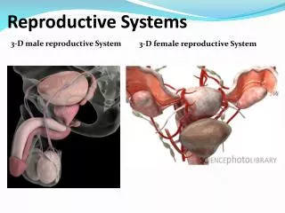
42.2 Reproductive System of Human Males • The male reproductive system produces hormones and sperm, which it delivers to a female reproductive tract • Male gonads (testes) • Primary reproductive organs in human males • Produce the male hormone testosterone • Sperm production begins at puberty
The Path of Sperm • Reproductive ducts • Seminal tubules produce immature sperm • Epididymis stores and matures sperm • Vas deferens carries sperm to ejaculatory duct • Ejaculatory duct connects to urethra in the penis • The penis contains spongy tissue which fills with blood during sexual excitement
Semen and Accessory Glands • Semen • Sperm and secretions from accessory glands (proteins, nutrients, ions, signaling molecules) • Accessory glands • Seminal vesicles secrete fructose-rich fluid (an energy source) and prostaglandins • Prostate gland produces alkaline secretions and prostaglandins • Bulbourethral glands secret lubricating mucus
Prostate and Testicular Problems • Prostate enlargement • Can be caused by inflammation, age, or prostate cancer – a leading cause of death • Constricts urethra, causing difficulty in urination • Diagnosed by blood tests, physical examination • Testicular cancer is relatively rare • Detected by self-examination
Ejaculatory Duct One pair of ducts that carry sperm to the penis Prostate Gland An exocrine gland that contributes some fluid to the semen Seminal Vesicle One of a pair of exocrine glands that contributes fructose-rich fluid to semen urinary bladder Urethra Duct with dual functions; channel for ejaculation of sperm during sexual arousal and for excretion of urine at other times Bulbourethral gland One of a pair of exocrine glands that secrete mucus Vas deferens One of a pair of ducts that carry sperm to the penis anus Epydidymis One of a pair of ducts in which sperm mature and are stored scrotum urethra Penis Male organ of sexual intercourse Testis One of a pair of gonads, packed with small, sperm-producing tubes (seminiferous tubules) and cells that secrete testosterone and other sex hormones cylinders of spongy tissue that swell with blood during an erection Fig. 42-4, p. 742
42.3 Sperm Formation • Seminiferous tubules in male testes continually produce new diploid germ cells (spermatogonia), which undergo meiosis to produce haploid male gametes (sperm) • Spermatogonium • Primary spermatocyte • Secondary spermatocyte • Spermatid • Immature sperm
vas deferens seminal vesicle prostate gland bulbourethral gland urethra penis epididymis seminiferous tubule testis Fig. 42-5a, p. 744
mitosis meiosis II meiosis I lumen immature sperm (haploid) Sertoli cell secondary spermatocyte early spermatids spermatogonium (diploid) primary spermatocyte late spermatid Fig. 42-5c, p. 744
Spermatozoan: a mature sperm Head with DNA and an enzyme cap Midpiece with mitochondria Flagellum for movement A Spermatozoan
head, with DNA and a cap of enzymes midpiece with mitochondria tail, with its core of microtubules Fig. 42-6, p. 745
Hormonal Control of Sperm Formation • The hypothalamus produces gonadotropin releasing hormone (GnRH) • GnRH causes the anterior pituitary to secrete follicle-stimulating hormone (FSH) and luteinizing hormone (LH)
Hormonal Control of Sperm Formation • LH causes Leydig cells between seminiferous tubules to produce testosterone • FSH causes Sertoli cells inside seminiferous tubules to produce growth factors and other molecular signals
Signaling Pathways in Sperm Formation:Inhibin and Negative Feedback Control
a Level of testosterone in blood decreases; the hypothalamus secretes GnRH, a releasing hormone. Hypothalamus f Elevated level of testosterone in blood inhibits secretion of GnRH. g High sperm count induces Sertoli cells to secrete inhibin, which inhibits secretion of GnRH and LH. Anterior Pituitary b GnRH stimulates secretion of LH, FSH from anterior lobe of pituitary. Testes d Sertoli cells bind FSH and testosterone, and function in spermatogenesis at puberty. c LH prompts Leydig cells in testes to produce and release testosterone. e Testosterone and secretions from Sertoli cells encourage sperm production. Fig. 42-7, p. 745
a Level of testosterone in blood decreases; the hypothalamus secretes GnRH, a releasing hormone. Hypothalamus f Elevated level of testosterone in blood inhibits secretion of GnRH. g High sperm count induces Sertoli cells to secrete inhibin, which inhibits secretion of GnRH and LH. Anterior Pituitary b GnRH stimulates secretion of LH, FSH from anterior lobe of pituitary. Testes d Sertoli cells bind FSH and testosterone, and function in spermatogenesis at puberty. c LH prompts Leydig cells in testes to produce and release testosterone. e Testosterone and secretions from Sertoli cells encourage sperm production. Stepped Art Fig. 42-7, p. 745
42.2-42.3 Key ConceptsMale Reproductive Function • A human male has a pair of testes that make sperm and secrete the sex hormone testosterone • Sperm mixes with secretions from other glands and leaves the body through ducts
42.4 Reproductive System of Human Females • The female reproductive system functions in the production of gametes and sex hormones • The system receives sperm, and has a chamber in which developing offspring are protected and nourished until birth
pelvic girdle uterus ovary urinary bladder vagina Fig. 42-8, p. 746
Components of the Female Reproductive System • Female gonads (ovaries) secrete sex hormones and produce immature eggs (oocytes) on a cyclic basis • Oocytes are released into an oviduct, where fertilization occurs, before entering the uterus, where the embryo develops • The vagina functions in intercourse and as the birth canal
Uterus Chamber in which embryo develops; its narrowed portion, the cervix, secretes mucus that helps sperm travel into the uterus and defends the embryo against many bacteria Oviduct One of a pair of ciliated channels through which oocytes are propelled from an ovary to the uterus; usual site of fertilization Ovary One of a pair of gonads that makes oocytes and sex hormones; during the course of a monthly cycle, releases hormones that stimulate maturation of an oocyte and prepares the lining of the uterus for a potential pregnancy Myometrium Thick muscle layers of uterus; stretch greatly during pregnancy Urinary bladder Endometrium Inner lining of the uterus into which a blastocyst implants itself; gets thicker and has increased blood supply during pregnancy; gives rise to maternal portion of placenta, an organ that metabolically supports embryonic and fetal development opening of cervix Urethra Clitoris Small organ responsive to sexual stimulation Labium Minora One of a pair of innermost thin, skin folds; part of the genitals anus Vagina Organ of sexual intercourse; also the birth canal Labium Majora One of a pair of outermost, fat- padded skin folds; part of the genitals Fig. 42-9, p. 746
Overview of the Menstrual Cycle • Menstrual cycle • A hormone-controlled estrous cycle in which a female is fertile only at certain times • Every 28 days, an oocyte matures and is released • The uterus prepares for pregnancy • If pregnancy does not occur, the endometrial lining of the uterus is shed (menstrual flow) • Menopause • Decline of hormone production and menstrual cycle