Download
1 / 88
880 likes | 885 Views
Reproductive Systems. Dr. Michael P. Gillespie. Sexual Reproduction. Process by which organisms produce offspring by making germ cells called gametes. The male gamete (sperm cell) unites with the female gamete (secondary oocyte). This is called fertilization.

E N D
Reproductive Systems Dr. Michael P. Gillespie
Sexual Reproduction • Process by which organisms produce offspring by making germ cells called gametes. • The male gamete (sperm cell) unites with the female gamete (secondary oocyte). This is called fertilization. • The resulting cell contains one set of chromosomes from each parent.
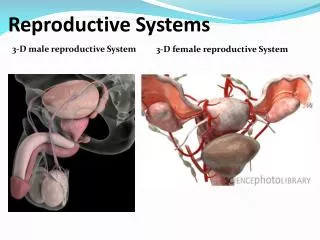
Reproductive Organs • Males and females have anatomically distinct reproductive organs that are adapted for producing gametes, facilitating fertilization, and in females sustaining the growth of the embryo and fetus.
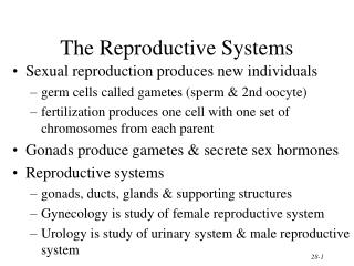
Reproductive Organs • The male and female reproductive organs can be grouped by function. • Gonads – testes in males and ovaries in females. • Produce gametes and secrete sex hormones.
Reproductive Organs • Ducts – store and transport gametes. • Accessory sex glands – produces substances that protect the gametes and facilitate movement. • Supporting structures: • Penis – facilitates delivery of gametes. • Uterus – facilitates delivery and joining of gametes. Facilitates growth of fetus during pregnancy.
Gynecology • Specialized branch of medicine concerned with diagnosis and treatment of diseases of the female reproductive system.
Urology • The study of the urinary system. • Urologists also diagnose and treat diseases of the male reproductive system.
Number Of Chromosomes • Human somatic cells contain 23 pairs of chromosomes, or a total of 46 chromosomes. • One member of each pair is inherited from each parent. • The members of each pair are called homologous chromosomes or homologs. They contain similar genes.
Number Of Chromosomes • Most of the homologs look the same with the exception of the sex chromosomes (designated X & Y). • Females carry to large X chromosomes. Males carry one large X and one small Y chromosome. • The other 22 pairs of chromosomes are called autosomes.
Number Of Chromosomes • Somatic cells contain 2 sets of chromosomes, so they are called diploid cells. • Geneticists use the symbol n to denote the number of different chromosomes in an organism. • In humans, n = 23. Diploid cells are 2n.
Meiosis • The reproductive cell division that occurs in the gonads. • It produces gametes in which the number of chromosomes is reduced by half. • Gametes contain a single set of 23 chromosomes and are called haploid cells. • Fertilization restores the diploid number of chromosomes.
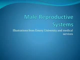
Male Reproductive System • The organs of the male reproductive system are the testes, ducts, accessory sex glands, and supporting structures including the scrotum and penis. • The testes produce sperm and secrete hormones. • A system of ducts transports and stores sperm. • Semen contains sperm plus the secretions provided by the accessory sex glands.
Functions Of The Male Reproductive System • Testes: produce sperm and the male sex hormone testosterone. • Ducts: transport, store, and assist in maturation of sperm. • Accessory sex glands: secrete most of the liquid portion of the sperm. • Penis: contains the urethra, a passageway for ejaculation and the excretion of urine.
Scrotum • The scrotum is the supporting structure for the testes. • Externally, the scrotum looks like a single pouch of skin separated into lateral portions by a median ridge of skin called the raphe. • Internally, the scrotal septum separates the scrotum into two sacs, each containing a single testis. • The septum contains muscle tissue called the dartos muscle.
Scrotum • When the dartos muscle contracts, it wrinkles the skin of the scrotum and elevates the testes. • The location of the scrotum and the contraction of its muscle fibers regulate the temperature of the testes.
Scrotum • Normal sperm production requires a temperature about 2-3 degrees C below core body temperature. • The scrotum is outside of the pelvic cavity and therefore maintains a lower temperature. • The cremaster muscle elevates the testes upon exposure to cold and during sexual arousal. This moves the testes closer to the pelvic cavity, where they can absorb body heat. The procedure is reversed in response to warmth.
Testes • The testes, or testicles, are paired oval glands. • A serous membrane called the tunica vaginalis partially covers the testes.
Testes • The tunica albuginea is internal to the tunica vaginalis and divides the testes into lobules. • The lobules contain 200-300 seminiferous tubules where sperm are produced. • 2 types of cells: • Spermatogenic cells – sperm forming cells. • Sertoli cells – supporting cells.
Testes • Spermatogenesis is the process by which sperm is produced. • Leydig cells lie between the seminiferous tubules and produce testosterone.
Cryptorchidism • A condition in which the testes do not descend into the scrotum. • It occurs in about 3% of full-term infants and 30% of premature infants.
Cryptorchidism • Untreated b/l cryptorchidism often results in sterility due to high temperatures. • The testes of about 80% of boys with cryptorchidism descend spontaneously within the 1st year of life. • Untreated, it results in a greater chance of testicular cancer.
Sperm • Spermatogenesis produces about 300 million sperm per day. • Once ejaculated, most sperm do not survive more than 48 hours in the female reproductive tract. • The sperm consists of a head with an acrosome (lysosomelike vesicle) and a nucleus with a haploid # of chromosomes (23).
Sperm • Enzymes within the acrosome aid in penetration of the sperm cell into the secondary oocyte. • The midpiece contains mitochondria. • The tail is a typical flagellum that propels the sperm cell.
Hormonal Control Of The Testes • Gonadotropin-releasing hormone (GnRH) – at puberty, the hypothalamus begins to release this hormone, which stimulates the release of luteinizing hormone (LH) and follicle-stimulating hormone (FSH). • LH stimulates Leydig cells to secrete testosterone. • FSH and testosterone stimulate spermatogenesis.
Ducts Of The Testis • Pressure generated by the fluid secreted by Sertoli cells pushes sperm and fluid along the lumen of the seminiferous tubules into the straight tubules. • The straight tubules lead to the rete testis and then into the efferent ducts. The efferent ducts empty into the ductus epididymis.
Epididymis • The epididymis is a comma-shaped organ that lies along the posterior border of each testis. • The ductus epididymis is the site where the sperm mature. • Sperm are stored here and peristaltic contraction propels them into the ductus (vas) deferens. • Sperm may remain here for a month or more.
Ductus Deferens • Within the tail of the epididymis, the ductus becomes less convoluted and is known as the ductus deferens or vas deferens. • Functionally, the ductus deferens stores sperm and conveys them toward the urethra by peristaltic contractions of the muscular coat. • Sperm that are not ejaculated are eventually reabsorbed.
Spermatic Cord • The spermatic cord is a supporting structure of the male reproductive system that ascends out of the scrotum. • The spermatic cord passes through the inguinal canal into the abdomen.
Spermatic Cord • The canal originates at the deep (abdominal) inguinal ring, a slitlike opening in the aponeurosis of the transversus abdominis muscle. • The canal ends at the superficial (subcutaneous) inguinal ring, an opening in the aponeurosis of the external oblique muscle.
Inguinal Hernias • The inguinal region is a weak area in the abdominal wall. • Consequently, it is often the site of an inguinal hernia – a rupture or separation of a portion of the inguinal area of the abdominal wall.
Inguinal Hernias • Indirect inguinal hernia – part of the small intestine protrudes through the deep inguinal ring and enters the scrotum. • Direct inguinal hernia – a portion of the small intestine pushes into the posterior wall of the inguinal canal causing a localized bulging in the wall of the canal.
Ejaculatory Ducts • The ejaculatory ducts are formed by the union of the duct from the seminal vesicle and the ampulla of the ductus deferens. • They eject sperm and seminal vesicle secretions into the urethra just before ejaculation (propulsion of semen from the urethra to the exterior).
Urethra • In males, the urethra is the shared terminal duct of the reproductive and urinary systems.
Accessory Sex Glands • The accessory sex glands secrete most of the liquid portion of the semen. • They include the seminal vesicles, the prostate, and the bulbourethral glands.
Functions Of The Accessory Sex Gland Secretions • Seminal vesicles – secrete alkaline, viscous fluid that helps neutralize the acid secretions of the female reproductive tract. Provides fructose for ATP production by sperm. Contributes to sperm motility, viability, and helps semen coagulate after ejaculation.
Functions Of The Accessory Sex Gland Secretions • Prostate – secretes a milky, slightly acidic fluid that helps semen coagulate after ejaculation and subsequently breaks down the clot. • Bulbourethral (Cowper’s) glands – secrete alkaline to neutralize the acidic environment of the urethra and mucous to lubricate the lining of the urethra and tip of the penis during intercourse.
Semen • Semen is a mixture of sperm and seminal fluid, a liquid that consists of the secretions of the seminiferous tubules, seminal vesicles, prostate, and bulbourethral glands. • Semen has a slightly alkaline ph of 7.2 – 7.7.
Semen • The prostatic secretions give the fluid a milky appearance, whereas fluids from the seminal vesicles and bulbourethral glands give it a sticky consistency. • Seminal fluid provides sperm with a transportation medium, nutrients, and neutralizes the hostile acidic environment of the male urethra and female vagina.
Penis • The penis contains the urethra and is a passageway for the ejaculation of semen and the secretion of urine. • It is cylindrical in shape and consists of a root, body, and glans penis. • Root of the penis is the attached (proximal) portion. It consists of the following: • Bulb of the penis (expanded portion of the base). • Crura of the penis (the 2 separated and tapered portions).
Penis • Body of the penis. • Glans of the penis – a slightly enlarged, acorn-shaped region (distal region). • The prepuce (foreskin) covers the glans in an uncircumcised penis. • The fundiform ligament and the suspensory ligament of the penis arise from the pubic symphisis and support the weight of the penis.
Erection • Upon sexual stimulation, the arteries supplying the penis dilate and large quantities of blood enter the sinuses. • The stimulation may be visual, tactile, auditory, olfactory, or imagined.