Download
1 / 50
610 likes | 1.12k Views
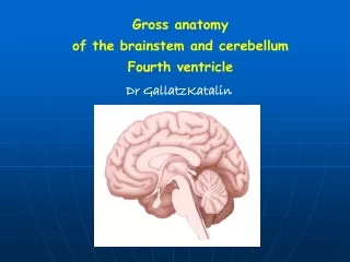
Chapter 3 Brainstem Anatomy, Topography, and Functional Levels. The brainstem: midbrain, pons, medulla oblongata. Midbrain. Tentorial notch. clivus. foramen magnum. posterior. anterior. 3 rd ventricle. Cerebral aqueduct. 4 th ventricle. central canal. Open part Closed part.

E N D
Chapter 3 Brainstem Anatomy, Topography, and Functional Levels
Midbrain Tentorial notch
clivus foramen magnum posterior anterior
3rd ventricle Cerebral aqueduct 4th ventricle central canal Open part Closed part
Medulla oblongata: Contains centers associated with: equilibrium audition deglutition coughing vomiting salivation tongue movements respiration circulation
Pons: Contains centers associated with: mastication eye movements facial expression blinking salivation equilibrium audition
Midbrain: Contains centers associated with: auditory reflexes visual reflexes pupillary reflexes
ventral surface of medulla Fig. 3-3
ventral surface of pons transverse bands Cerebellar angle junction Fig. 3-3
Basilar artery Vertebral artery
ventral surface of midbrain (anterior part of cerebral peduncle) Fig. 3-3
dorsal surface of closed medulla Fig. 3-4
Floor of 4th ventricle: (see Fig. 3-4) caudal part is formed by posterior surface of rostral medulla
Floor of 4th ventricle: rostral part is formed by posterior surface of pons
motor structures are more medial; sensory structures are more lateral
Fig. 5-3 Motor cranial nerves
ventral surface of medulla (cranial rootlets) Fig. 3-3
ventral surface of pons transverse bands Cerebellar angle junction Fig. 3-3
ventral surface of midbrain CN’s I and II connect to the forebrain (anterior part of cerebral peduncle) Fig. 3-3
Myelin-stained brainstem cross sections: Rostral part of closed medulla (at dorsal column nuclei)
{ the dorsal column nuclei Fig. 3-6 Rostral part of closed medulla
Caudal part of open medulla (at hypoglassal and vagal nuclei)
hypoglossal nucleus nucleus ambiguus Fig. 3-7 Caudal part of open medulla (at hypoglossal and vagal nuclei)
acoustic tubercle (opens into subarachnoid space) Inferior cerebellar peduncle Fig. 3-8 Rostral part of open medulla (at lateral aperture)
Facial nucleus Fig. 3-9 Caudal pons (at abducens and facial nuclei)
(emergence) Rostral pons (at decussation of trochlear nerves)
Trochlear nucleus Caudal midbrain (at inferior colliculus)
CNIII fibers Rostral midbrain (at superior colliculus)
corpora quadrigemina (for reflex movements of eyes, head, and neck)
Inputs and outputs of the Brainstem Reticular Formation Fig. 20-2
Functions of the reticular formation: 1. Integrates of cranial nerve reflexes 2. Helps in conduction and modulation of slow pain 3. Influences voluntary movements 4. Regulates autonomic nuclei 5. Is associated with diffuse modulating systems 6. Integrates basic functions (respiration, sleep) 7. Activates the cerebral cortex * * Ascending Reticular Activating System Fig. 20-9
Chapter 3 know the location of the brainstem and the locations where it is continuous with the forebrain and the spinal cord know the functional centers contained within the midbrain, pons, and medulla know the locations of the tectum and tegmentum of the brainstem know the anterior surface features of the medulla know the anterior surface features of the pons know the anterior surface features of the midbrain know the posterior surface features of the closed medulla know the parts of the brainstem that form the floor of the 4th ventricle know the posterior surface features seen in the floor of the 4th ventricle know the posterior surface features of the midbrain know the points of attachment or emergence of the cranial nerves III-XII know the cross section levels of the brainstem where the nuclei of the motor cranial nerves are found