Download
1 / 28
280 likes | 615 Views
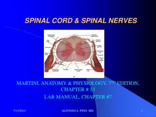
CNS – The Spinal Cord, Spinal Nerves & Spinal Reflexes. Spinal Cord & Spinal Nerves. Spinal cord Truly the pathway between body and mind Conducts impulses to and from the brain Carries out spinal reflexes Spinal nerves 31 pairs All are mixed nerves. Mixed Nerves?. Dorsal roots

E N D
Spinal Cord & Spinal Nerves • Spinal cord • Truly the pathway between body and mind • Conducts impulses to and from the brain • Carries out spinal reflexes • Spinal nerves • 31 pairs • All are mixed nerves
Mixed Nerves? • Dorsal roots • Ganglion containing axons of sensory neurons (afferent) • Ventral roots • All motor fibers (axons) leave the cord by the ventral roots (efferent)
General Organization • Highly organized, very efficient
Spinal Reflexes • Rapid, automatic nerve responses triggered by specific stimuli • Controlled by spinal cord alone; not the brain
The Adult Spinal Cord • About 18 inches (45 cm) long • 1/2 inch (14 mm) wide • Ends between vertebrae L1 and L2
Bilateral Symmetry • Grooves divide the spinal cord into left and right • Posterior median sulcus: • Shallow • Anterior median fissure: • deeper groove on anterior side
Enlargements of the Spinal Cord • Caused by: • amount of gray matter in segment • involvement with sensory and motor nerves of limbs • Cervical enlargement: • nerves of shoulders and upper limbs • Lumbar enlargement: • nerves of pelvis and lower limbs
The Distal End • Conus medullaris: • thin, conical spinal cord below lumbar enlargement • Filum terminale: • thin thread of fibrous tissue at end of conus medullaris • attaches to coccygeal ligament
The Meninges • Specialized membranes isolate spinal cord from surroundings • Spinal meninges: • protect spinal cord • carry blood supply • continuous with cranial meninges • Meningitis: • viral or bacterial infection of meninges
The 3 Meningeal Layers • Dura mater: • outer layer of spinal cord • Arachnoid mater: • middle meningeal layer • Pia mater: • inner meningeal layer
Spinal Dura Mater • Are tough and fibrous • Cranially: • fuses with periosteum of occipital bone • is continuous with cranial dura mater • Caudally: • tapers to dense cord of collagen fibers • joins filum terminale in coccygeal ligament
The Epidural Space • Between spinal dura mater and walls of vertebral canal • Contains loose connective and adipose tissue • Anesthetic injection site
Arachnoid Mater • Middle meningeal layer • Arachnoid membrane: • simple squamous epithelia • covers arachnoid mater
Inter-Layer Spaces • filled with cerebrospinal fluid(CSF)
Cerebrospinal Fluid (CSF) • Is found in subarachnoid space • Carries dissolved gases, nutrients, and wastes • Spinal tap: • withdraws CSF
Pia Mater • Is the innermost meningeal layer • Is a mesh of collagen and elastic fibers • Is bound to underlying neural tissue
Consists of…. • White matter • Gray matter • Gray horns • Posterior gray horns • contain somatic and visceral sensory nuclei • Anterior gray horns • contain somatic motor nuclei • Lateral gray horns • are in thoracic and lumbar segments • contain visceral motor nuclei
Spinal cord is so highly organized: • it is possible to predict results of injuries to specific areas
Spinal Nerves • The roots unite into the spinal nerve • Spinal nervesexit through intervertebral foramen • Splitintobranches, or rami.
Dermatomes Specific bilateral region of the skin surface monitored by a single pair of spinal nerves
Nerve Plexuses • Plexus = “braid” • Nerves supplying the limbs form plexuses when they leave the spinal cord • Cervical plexus • Brachial plexus • Lumbar plexus • Sacral plexus