Download
1 / 25
260 likes | 312 Views
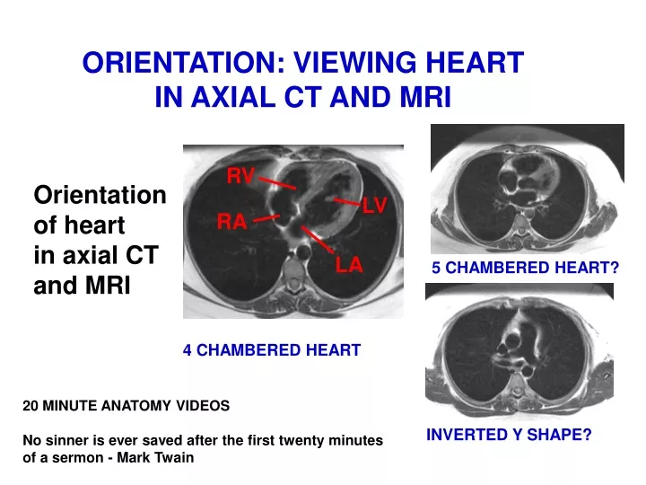
ORIENTATION: VIEWING HEART IN AXIAL CT AND MRI. Orientation of heart in axial CT and MRI. RV. LV. RA. LA. 5 CHAMBERED HEART?. 4 CHAMBERED HEART. 20 MINUTE ANATOMY VIDEOS No sinner is ever saved after the first twenty minutes of a sermon - Mark Twain. INVERTED Y SHAPE?.

E N D
ORIENTATION: VIEWING HEART IN AXIAL CT AND MRI Orientation of heart in axial CT and MRI RV LV RA LA 5 CHAMBERED HEART? 4 CHAMBERED HEART 20 MINUTE ANATOMY VIDEOS No sinner is ever saved after the first twenty minutes of a sermon - Mark Twain INVERTED Y SHAPE?
ORIENTATION: THORACIC CONTENTS ANTERIOR VIEW Esophagus Trachea Left Int. Jugular V. Subclavian V. R. Brachiocephalic V. AORTA Sup. Vena Cava Structures at Root of Lung Pulmonary Trunk diaphragm L R
POSTERIOR VIEW INCLUDING TRACHEA, ESOPHAGUS Esophagus, Thoracic duct 1) Arch of Aorta 2a) Trachea (bifurcation) 2) Pulmonary Arteries Order similar to root of Lung 3) Pulmonary Veins diaphragm IVC R L
HEART IS ON LEFT: REASON: ROTATED SO LEFT ATRIUM IS IN MIDLINE Left Atrium - receives Pulmonary Veins symmetrically from both sides Right Ventricle - anterior Left Pulmonary Veins LA Right Pulmonary Veins RV
POSTERIOR VIEW: KNOW SEQUENCE SVC 1) Arch of Aorta 2) Pulmonary Arteries Pulmonary Veins 3) Pulmonary Veins IVC
ANTERIOR VIEW AT TOP (SUPERIORLY) - VEIN IN FRONT OF ARTERIES Left Brachiocephalic Vein Brachiocephalic Trunk Left Common Carotid A Left Subclavian A Right Brachiocephalic Vein SVC Arch of Aorta ANTERIOR VIEW Reason: All venous blood drains to Right Atrium
ANTERIOR VIEW PULMONARY TRUNK - arises from Right Ventricle; crosses anterior to Ascending Aorta; then branches Arch of Aorta Ascending Aorta Pulmonary Trunk - from Conus Arteriosus RV
CONUS ARTERIOSUS EXTENDS TOWARD LEFT; THEN PULMONARY TRUNK BIFURCATES RA Y LA PULMONARY TRUNK ON LEFT IN AXIAL SECTIONS Conus Arteriosus RV LV
AORTA ARISES IN MIDDLE OF HEART RA LA AXIAL SECTIONS LOOK LIKE 5 CHAMBERED HEART RV LV
POSTERIOR VIEW: KNOW SEQUENCE SVC 1) Arch of Aorta 2) Pulmonary Arteries Pulmonary Veins 3) Pulmonary Veins R L IVC
ORIENTATION: VIEWING HEART IN AXIAL CT AND MRI RV Orientation of heart in axial CT and MRI LV RA LA
CT, MRI VIEW HEART FROM BELOW LV RV LA RA * - AORTA IN CENTER OF HEART L IMAGE FLIPPED VERTICALLY R
START AT DIAPHRAGM Diaphragm Inferior vena cava Muscles of ventricles Esophagus Azygous vein Thoracic (descending) aorta R L
Right ventricle Interventricular septum Inferior vena cava Left ventricle Esophagus Azygous vein Thoracic (descending) aorta R L 2 CHAMBERED HEART - ventricles only
Right ventricle Right atrium Left ventricle Left atrium Esophagus Thoracic (descending) aorta R L 4 CHAMBERED HEART!
Right ventricle Right atrioventricular orifice Interventricular septum Right atrium Left ventricle Right pulmonary vein Left atrioventricular orifice LA Left atrium Thoracic (descending) aorta Azygous vein Left pulmonary vein R L 4 CHAMBERED HEART!
Right ventricle * - AORTA - arising from Vestibule Right atrium Left ventricle Left atrium Right pulmonary vein Azygous vein Left pulmonary vein Esophagus R Thoracic (descending) aorta L 5 CHAMBERED HEART?
Right ventricle Superior vena cava Ascending aorta RV Azygous vein Left main bronchus R L RIGHT VENTRICLE EXTENDS TO LEFT
Right ventricle to Pulmonary Trunk Ascending aorta Superior vena cava Left pulmonary artery Y Y Right pulmonary artery Thoracic (descending) aorta Carina at tracheal bifurcation R L PULMONARY TRUNK BRANCHES
Arch of aorta Superior vena cava Trachea Esophagus R L
Superior vena cava Left subclavian artery Trachea Scapula Spinal cord Esophagus R L
Left brachiocephalic vein Right brachiocephalic vein Right lung Brachiocephalic trunk Left common carotid R L Left subclavian artery Trachea Esophagus LEFT BRACHIOCEPHALIC VEIN CROSSES ANTERIORLY
Summary - Orientation of heart in CT and MRI is complex; rarely appears to have 4 chambers. 1. Position and Orientation of heart - Heart is rotated and tilted so Right Ventricle is anterior and inferior; this makes functional sense; as a result, Left Atrium is located in midline and receives blood symmetrically from Pulmonary Veins (low pressure). 2. Sequence of Structures Inferior to Superior - Pulmonary Veins, Pulmonary artery, Arch of Aorta. 3. Pulmonary artery - arises from Conus arteriosus (Infundibulum) of Right Ventricle; ascends toward left and crosses anterior to Aorta; Pulmonary artery then bifurcates (appears Y shaped in axial CT and MRI). 4. Aorta - arises from Left Ventricle and first courses in middle of heart (Heart appears to have 5 chambers in axial CT and MRI at this level). 5. Order of Structures in Superior Mediastinum - Veins (R and L Brachiocephalic) are anterior to Arteries (Brachiocephalic, L Common Carotid, L Subclavian) arising from Arch of Aorta.
REMEMBER: ANATOMICAL DETAILS ARE COMPLEX AND DIFFICULT TO LEARN BUT PATIENTS CAN DIE FROM 'MINOR DETAILS' OF ANATOMY.