Download
1 / 50
500 likes | 546 Views
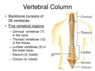
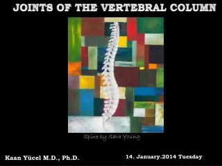
Dr. Nabil Khouri MD, MSc, Ph.D. The Vertebral column. Vertebral column. 7. 12. 5. 5. 4. 33 Vertebrae Inter-vertebral disc Form 1/4 of its length. Vertebra Column Distributed as follows. 7 cervical vertebrae – neck region 12 thoracic vertebrae – posterior to the thoracic cavity

E N D
Dr. Nabil Khouri MD, MSc, Ph.D The Vertebral column
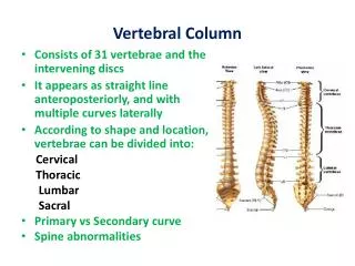
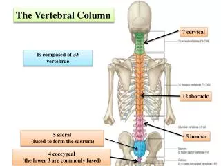
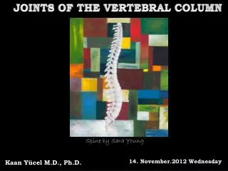
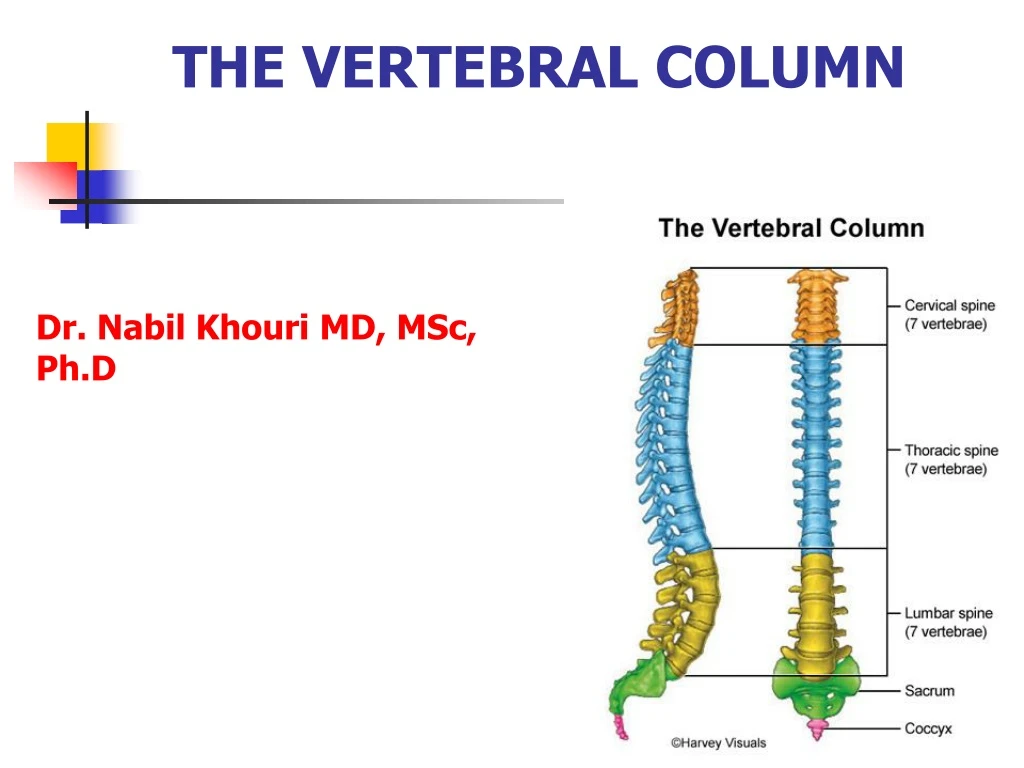
Vertebral column 7 12 5 5 4 33 Vertebrae Inter-vertebral disc Form 1/4 of its length
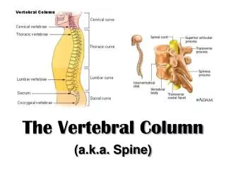
Vertebra ColumnDistributed as follows • 7 cervical vertebrae – neck region • 12 thoracic vertebrae – posterior to the thoracic cavity • 5 lumbar vertebrae – support the lower back. • 1 sacrum – consists of 5 fused sacral vertebrae. • 1 coccyx – consists of 4 fused coccygeal vertebrae.
Curvature of vertebral column Thoracic and sacral (primary) Concave anterior. Develop during fetal period, deference between ant and post Thickness of the vertebra. Cervical and lumbar : Concave posterior, develop during the fetal period, due to deference in IV disc thickness - Cervical - infant hold head - Lumber - infant walk and assume upright position, prominent in female.
Sagittal Plane Curves • Primary Curves • Secondary Curves
Line of gravity Regions of the Spine Auricle of the ear Odontoid Body of C7 Anterior to thoracic spine Posterior to L3 Mid femoral heads
Abnormal Vertebral column curvature Kyphosis: abnormal increase in thoracic curv. Erosion of anterior vertebral part. Lordosis: (hollow back) anterior rotation of pelvis. Abnormal increase in lumber curvature (pregnancy) Scoliosis: (Crooked or curved back) abnormal lateral curvature and rotation of the back (appears between ages of 10-15)
General Structure of Typical Vertebrae Body or centrum – disc-shaped, weight-bearing region Vertebral arch – composed of Pedicles and Laminae that along with the centrum, encloses the Vertebral Foramen Vertebral foramina – make up the vertebral canal through which the spinal cord passes
Vertebral Arch 2 short, thick processes called the pedicles, project posteriorly from the vertebrae body to unite with the flat laminae, to form the vertebrae arch. Vertebral arch extend posteriorly from the body of the vertebra, together with the body and vertebral arch surround the spinal cord by forming the vertebral foramen. Vertebral foramen contain spinal cord, adipose tissue and areolar connective tissue and blood vessels.
General Structure of typical Vertebrae cont. 7 processes arise from the vertebral arch. One Spinous process Is a projection posterior and usually it is oriented downward. Transverse processes: Are two projections laterally
Cont.. • The remaining 4 processes forms joints with other vertebrae above and below. • 2 superior articular processes articulate with the 2 inferior articular processes of vertebrae above them. • The articulating surfaces of articular processes called facets. • Articulation between the bodies and articular facets of successive vertebrae are called intervertebral joint.
Inter-vertebral foramina – lateral openings formed from notched areas on the superior and inferior borders of adjacent pedicles
Intervertebral Discs • Intervertebral fibrocartilage. • Lie between adjacent vertebrae in the spine. • Each disc forms a cartilaginous joint to allow slight movement of the vertebrae • Acts as a ligaments to hold the vertebrae together.
Cervical Vertebrae • C1-C7 • C1, C2, C7 = Atypical , C2 – C6 = Typical • The bodies are smaller than thoracic vertebrae. • Vertebral arches are larger. • Have one vertebral foramina, and two transverse foramina. • Vertebral foramina of cervical vertebrae are the largest in the spinal column because they house the cervical enlargement of the spinal cord.
Cont… • Each cervical transverse processes contain a transverse foramen through which the vertebral artery, vein and nerve pass. • Spinous processes of C2-C6 are often bifid – split into two parts. • The first two cervical vertebra considerably from others. • First cervical vertebrae (C1) called atlas, and second cervical vertebrae (C2) called axis.
Cervical 1 (Atlas) • The atlas is a ring of bone with anterior and posterior arches and large lateral masses. • Lacks a body and a spinous process. • Large vertebral foramen (triangular) • The superior surface of the lateral masses called superior articular facets are concave. • Superior articular facets articulate with occipital condyle of occipital bone to form atlanto – occipital joint.
The Atlas (C1) Anterior Tubercle Articular Facet for Dens Transverse Process Superior Articular Facet Transverse Foramen Lateral Mass Lamina Posterior Tubercle Superior View
Cont… • These articulation permits the movement for “yes”. • Inferior surface of the lateral masses called inferior articular facets,articulate with axis vertebrae to form atlanto-axial joint. • Transverse process and transverse foramina of the atlas are quiet large. • The large vertebral foramina divide into 2 foramina by the transverse ligament; larger posterior foramina (spinal cord) and smaller anterior foramina (dens)
Cervical 2 (Axis) • Atypical cervical vertebra • Have a body • Peglike process called dens or odontoid process projects up through anterior portion of the vertebral foramen of the atlas. • The dens makes a pivot on which the atlas and head rotate, as in moving the head to signify “ NO”.
The Axis (C2) Lateral Mass Odontoid Process (Dens) Body Superior Articular Facet Inferior Articular Facet Spinous Process Transverse Process Anterior View Posterior View
Cervical 2 (Axis) Hermizan Halihanafiah
Atlanto-Axial Joint Hermizan Halihanafiah
Pivot Joint Cont… • This arrangement allow side to side rotation of the head. • The articulation formed between the anterior arch (facet) of the atlas and dens of the axis, and between their articulation facets (inferior and superior articular facets) called the atlanto-axial joint. • Movements: Rotation Around one Axes
Cervical 7 (C7) • Called the vertebra prominents. • Has single large spinous process that can be felt at the base of the neck. • Spinous process is not bifid. • Body is larger. • Pedicles are directed more posteriorly than laterally. • Inferior articular facets face more anteriorly than downwards. • Vertebral foramen, generally smaller than other cervical vertebrae.
Cervical 7 (C7) Spinous process not bifid, large project posteriorly Vertebral Prominents Hermizan Halihanafiah
Cervical Vertebra (C7) Its has a long and prominent spinous process. Its thick, nearly horizontal, not bifurcated. Foramen transversorium may be as large as that in the other cervical vertebrae On the left side it occasionally gives passage to the vertebral artery; more frequently the vertebral vein transverses it on both sides; but the usual arrangement is for both artery and vein to pass in front of the transverse process, and not through the foramen. Sometimes the anterior root of the transverse process attains a large size and exists as a separate bone, which is known as a cervical rib.
Cervical 7 (C7) Hermizan Halihanafiah
Thoracic Vertebrae There are twelve vertebrae (T1-T12) all of which articulate with ribs. Major markings include two demi-facets on the heart-shaped body for the head of the rib. Circular vertebral foramen, transverse processes with articular costal facets for the rib tubercles. Long biffed spinous process that is inclined downward. The location of the articulate facets prevent flexion and extension, but allow rotation of this area of the spine. Superior articular processes are oriented backward and laterally.
The five lumbar vertebrae (L1-L5) are located in the small region of the back and have an enhanced weight-bearing function. Body is large and kidney-shaped. They have short, thick pedicles and lamina. Flat hatchet-shaped spinous processes. Triangular-shaped vertebral foramen. Orientation of the sup. articular facets face medially to lock the lumbar vertebrae together to provide stability Lumbar Vertebrae
Lumbar Vertebrae, L1-L5 • Body - L1 to L5 progressive increase in mass • Pedicles - longer and wider than thoracic; oval shaped • Spinous processes - horizontal, square shaped • Transverse processes - smaller than in thoracic region • Intervertebral foramen - large, but with increased incidence of nerve root compression
Sacrum and Coccyx The sacrum Consists of five fused vertebrae (S1-S5), which shape the posterior wall of the pelvis It articulates with L5 superiorly, and with the auricular surfaces of the hip bones Major markings include the sacral promontory, transverse lines, alae, dorsal sacral foramina, sacral canal, and sacral hiatus
S1 S2 S3 S4 S5 1 2 3 4 Coccyx • Coccyx (Tailbone) • The coccyx is made up of four (in some cases three to five) fused vertebrae that articulate superiorly with the sacrum
Disc Problems Slipped (degeneration) disc vs. herniated disc Most common sites for disc problems: C5 - C6 L4 - L5 L5 - S1 Lumbago Laminectomy ( IS a surgical removal vertebral arch by shaving laminae to access disc)