Download
1 / 114
1.15k likes | 1.22k Views
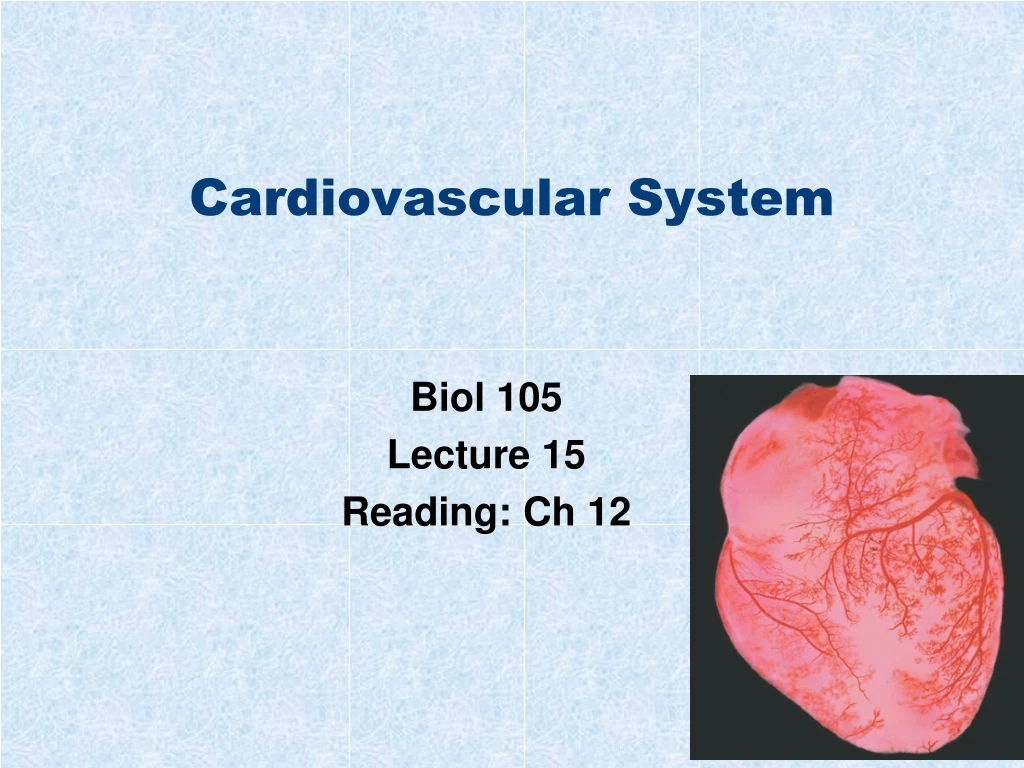
Cardiovascular System. Biol 105 Lecture 15 Reading: Ch 12. Outline. Functions of cardiovascular system Components of the cardiovascular system The heart Regulation of the heartbeat ECG/EKG Blood pressure Circulatory circuits Cardiovascular diseases Lymphatic system. Cardiovascular.

E N D
Cardiovascular System Biol 105 Lecture 15 Reading: Ch 12
Outline • Functions of cardiovascular system • Components of the cardiovascular system • The heart • Regulation of the heartbeat • ECG/EKG • Blood pressure • Circulatory circuits • Cardiovascular diseases • Lymphatic system
Cardiovascular • Function of the cardiovascular system is to transportblood containing: • Nutrients • Waste • Hormones • Immune cells • Oxygen
Cardiovascular system • Cardiovascular System consists of three components: • Blood • The heart, which pumps blood. • The blood vessels, through which blood flows. 5-2
The Cardiovascular System - Veins Veins • Carry blood back to theheart Jugular veins • Carry blood from headto the heart Superior vena cava • Carries blood from theupper body back to theheart Pulmonary veins • Carry oxygenated bloodfrom the lungs to theheart Renal vein • Carries blood from thekidney to the heart Inferior vena cava • Carries blood from thelower body back to theheart Iliac vein • Carries blood from thepelvic organs andabdominal wallback to the heart Radial vein • Carries blood from thehand back to the heart Femoral vein • Carries blood from thethigh and inner kneeback to the heart Figure 12.1 (1 of 2)
The Cardiovascular System - Arteries Arteries • Carry blood away fromheart Aorta • Delivers blood to thebody tissues Carotid arteries • Deliver blood to the headand the brain Pulmonary arteries • Deliver oxygen-poorblood to the lungs Coronary arteries • Deliver blood to theheart muscle cells Renal artery • Delivers blood to thekidney Iliac artery • Delivers blood topelvic organs andabdominal wall Radial artery • Delivers blood tothe hand Femoral artery • Delivers blood tothigh and inner knee Figure 12.1 (2 of 2)
The Heart and Lungs Figure 12.7b
Blood vessels • Blood vessels are lined with epithelial cells • They have a layer of smooth muscles that contract or dilate the vessels • Blood vessels are covered with a layer of connective tissue • Inside the vessels is called the lumen.
Vasoconstriction and Vasodilation • Vasoconstriction • When muscle contracts and the diameter of the lumen narrows, reducing blood flow • Vasodilation • When muscle relaxes and the diameter of the lumen increases, increasing blood flow
The Blood Vessels • Arteries • Arterioles • Capillaries • Venules • Veins
The Blood Vessels – Arteries and Veins • Arteries - Always carry blood away from the heart and usually carry O2-rich blood. • Veins - Always returns blood to the heart and usually carry O2-poor blood. 5-4
The Blood Vessels – Arterioles and Venules • Arteries break down into smaller vessels called arterioles, bringing O2, water, and nutrients to the tissues • Arterioles break down into small vessels called capillaries • Blood leaves the capillaries and enters venules • Venules take CO2, water, and wastes away from the tissues. • Venules join together to form veins. 5-4
The Blood Vessels – Arterioles • There are sphincter muscles that contract to reduce blood flow to the capillaries • or they dilate to increase blood flow to the capillaries. 5-4
Capillaries • Small vessels are called capillaries • It is here that components (O2, CO2, nutrients, waste) can pass from the blood vessels to other tissues • Capillaries do not have a smooth muscle layer
Can gas freely pass through the plasma membrane? • True • False
Capillaries • The RBCs stay in the blood vessels but the oxygen leaves the RBCs and the capillaries and goes into the tissues. • The oxygen leaves the capillaries because there is a gradient – there is more oxygen in the capillaries than in the tissues.
Capillaries • Blood flow in capillaries is slow. • This is important because it allows time for the exchange of substances between the blood and surrounding tissues. 5-18
The Blood Vessels Figure 12.2
Blood Vessels Figure 12.4b
Blood Vessels Figure 12.4a
Capillaries Figure 12.3c
Capillaries To tissue cells Slit between cells Capillary cell Nucleus Red blood cell (a) Substances are exchanged between the blood and tissuefluid across the plasma membrane of the capillary or throughslits between capillary cells. Figure 12.3a
Capillaries Figure 12.3b
Do RBCs leave the capillaries? • Yes • No
Pressures and Their Effect on Capillaries • At the arterial end of the capillaries blood pressure forces fluid out of the capillary and into the tissue • At the venous end, osmotic pressure draws fluid back into the vessel from the tissue • Diffusion is the pressure that draws gasses across the capillary
The Blood Vessels • Arteries • Aorta — largest artery. • Arterioles — smallest arteries (whether constricted or dilated affects blood pressure). • Veins • Vena cava — largest veins in the body. • Venules — smallest veins. 5-4
The Heart Aorta Pulmonaryveins Superiorvena cava Pulmonarytrunk Rightcoronaryvein Left coronaryartery Leftcoronaryvein Rightcoronaryartery Inferiorvenacava (a) Figure 12.10a
The Heart Superior venacava Aorta Left pulmonaryarteries Rightpulmonaryarteries Pulmonary trunk Pulmonarysemilunar valve Left pulmonary veins Right atrium Left atrium Rightpulmonaryveins Aortic semilunar valve(hidden from view) Leftatrioventricularvalve (mitral valve) Rightatrioventricularvalve(tricuspid valve) Left ventricle Chordae tendineae Myocardium Rightventricle Endocardium Pericardium Inferior vena cava (d) Septum Figure 12.7d
The Heart • The heart is composed of four chambers and lies almost in the center of the thoracic cavity. • Two atria—thin-walled upper chambers that serve as reservoirs for blood. • Two ventricles—thick-walled lower chambers powering the pulmonary and systemic circuits. • Septum—separates right and left sides of the heart. 5-7
The Heart • There are valves which keep blood flowing forward: • Two atrioventrical valves (AV) — between atria & ventricles, making a “LUB” sound when closing. • Two semilunar vales (SL) — base of major arteries making a “DUB” sound when closing. 5-7
The Heart Valves Figure 12.8
The Heart • Pericardium — thick membranous sac surrounding the heart (secretes serous fluid). • Myocardium — consists of cardiac muscle tissue, which contracts to pump blood. • The interior of the heart is lined by endocardium 5-7
The Heart Figure 12.7a
The Heart Oxygen-richblood(to body) Oxygen-poorblood(to lungs) Oxygen-poorblood(from body cells) Oxygen-richblood (from lungs) (c) Figure 12.7c
Path of Blood Through Heart • Superior and Inferior vena cava (O2-poor) Right Atrium. • R Atrium Tricuspid AV valve Right Ventricle. • R Ventricle Pulmonary SL valve Pulmonary Arteries Lungs. • Pulmonary veins (O2-rich) Left Atrium. • L Atrium Mitral AV valve Left Ventricle. • L Ventricle Aortic SL valve Aorta rest of the body tissues. 5-9
Cardiac Cycle • Cardiac cycle - one complete heart beat where both atria contract simultaneously (at the same time) followed by both ventricles contracting simultaneously. • a. Systole - when ventricles contract and pump blood out of the heart. • b. Diastole - when ventricles relax and receive blood from atria. 5-11
Heartbeat regulation - Intrinsic • Intrinsic Control: • Sinoatrial node (SA) (pacemaker)—initiates the heartbeat and causes the atria to contract. • Atrioventricular node (AV) - causes the ventricles to contract.
Heartbeat regulation - Intrinsic • The AV node relays the message to the ventricles using bundles of specialized muscle cells = atrioventricular bundle • The bundle divides into smaller bundles of specialized cardiac muscle cells called Purkinje fibers
When the ventricles contract, which valves are closed? • AV valves • SL valves
Heartbeat regulation - Extrinsic • Extrinsic Control of Heartbeat - the autonomic nervous system and hormones can modify the rate of the heartbeat.
Which part of the autonomic NS controls the heart most of the time? • Sympathetic • Parasympathetic
Regulation of the Heartbeat Figure 12.12 (1 of 5)
Regulation of the Heartbeat Figure 12.12 (2 of 5)
Regulation of the Heartbeat Figure 12.12 (3 of 5)
Regulation of the Heartbeat Figure 12.12 (4 of 5)
Regulation of the Heartbeat Figure 12.12 (5 of 5)