Download
1 / 28
290 likes | 324 Views
Cardiovascular System. By: Kayla Sood & Cynthia Amador P.4. What is the C ardiovascular System ?. Why is the cardiovascular system important to us?. Carries oxygen, nutrients, hormones, proteins, electrolytes, gases to cells. Transports toxins and carbon dioxide away from cells.

E N D
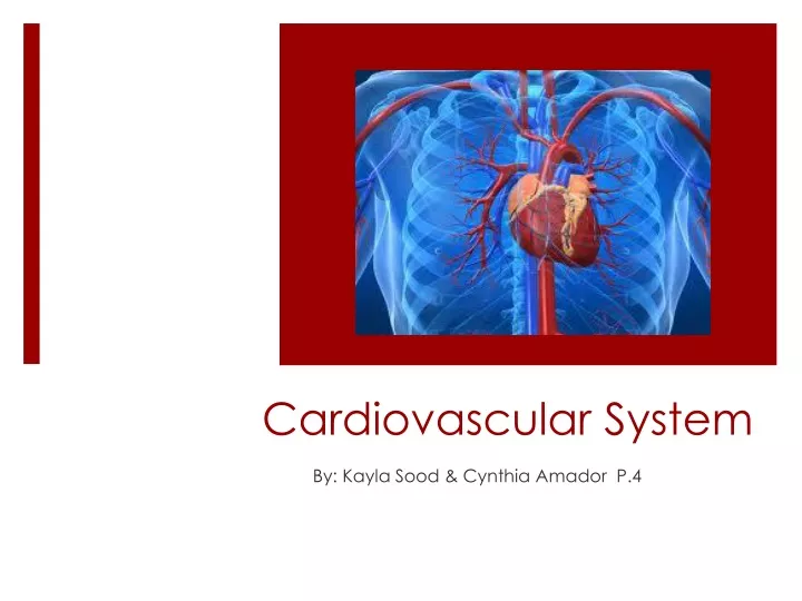
Cardiovascular System By: Kayla Sood & Cynthia Amador P.4
Why is the cardiovascular system important to us? • Carries oxygen, nutrients, hormones, proteins, electrolytes, gases to cells. • Transports toxins and carbon dioxide away from cells. • Necessary to stay alive, duhhh!
Heart Chambers and Valves The heart consists of 4 hallow chambers and 4 valves, that make the process of blood circulation throughout the body possible.
Heart chambers and valves(continued…) Atria- upper chambers • thin walls • Receives blood returning to heart Ventricle- lower chambers • Forces blood out heart, into arteries Aorta- carries blood from heart to rest of body except lungs Interatrial Septum • Separates right and left atrium Interventricular Septum • Separates right and left ventricles
4 Chambers Left atrium • Receives oxygen rich-blood from lungs • Pumps to left ventricle Left ventricle • Pumps oxygen rich-blood to aorta then out to the body Right atrium • Receives oxygen • Pumps to right ventricle Right ventricle • Pumps oxygen poor-blood to lungs
4 Valves 2 Semilunar • Mitral Valve (bicuspid valve) • Located on left side • Tricuspid Valve • Located between upper atria and lower ventricles on right side 2 Atrioventricular • Aortic Valve • Located on the left side • Pulmonary Valve • Located in arteries leaving the heart on right side
Purposes of the 4 major valves Mitral and tricuspid • Control blood flow from atria to ventricle Aortic and pulmonary • Control blood flow out of the ventricles As LV contracts, opens and allows blood to leave; closes and prevents back flow of blood in LV When ventricles contract, prevents back flow of blood in LA and LV Allows blood flow from RA into RV, prevents blood moving in wrong direction Prevents backflow of blood into RV and opens as it contracts
Coverings of the Heart The heart consist of two major layers: Heart is enclosed in a double-walled sac: pericardium The loose fitting superficial part of this sac: fibrous pericardium. The fibrous pericardium: protects heart holds together surrounding structures prevents overfilling heart with blood Deeper part of fibrous pericardium is the serous pericardium…
Coverings of the heart (continued…) Serous Pericardium: a thin, two-layer membrane that forms a closed sac around the heart. Parietal layer of serous pericardium lines internal surface of the fibrous pericardium. At superior edges of heart, parietal layer attaches to large arteries exiting the heart. Then turns inferiorly and continues over the external heart surface as the visceral layer, also called theepicardium, *which is an essential part of the heart wall.
Coverings of the heart (continued…) Layers of the Heart Wall The heart wall is composed of three layers: • Epicardium • myocardium • endocardium • Epicardium(superficial layer) is the visceral layer of the serous pericardium. • Thin layer of CT and fat • Myocardium(middle layer)composed mainly of cardiac muscle cells (cardiomyocytes) and forms the bulk of the heart. • CT fibers form a dense network called the fibrous cardiac skeleton that support structure of the myocardium & cardiac muscle fibers. • Inner layer:Endocardium made up of endothelial & consist of thin, smooth membrane, lines inside chambers of heart, located under myocardium.
Coverings of the heart (continued…) pericardial cavity: Between the parietal and visceral layers;contains a film of serous fluid. The serous membranes are lubricated by the serous fluid Allows for gliding past each other, allowing the heart to work in a friction-free environment.
Blood vessels Arteries: Vessels that transports blood away form the heart & to capillaries Capillaries: small blood vessel that connects an arteriole to a venule; thin and fragile Veins: vessels that carry blood towards the heart 5 major vessel that go to and leave the heart: • Superior vena cava • Inferior vena cava • Pulmonary artery • Pulmonary vein • aorta
Blood vessels(continued…) • Vena cava: veins that return deoxygenated blood from circulation & body and empty into the right atrium • Superior: transports deoxygenated blood from upper extremities (head, neck etc.) • Inferior: transports deoxygenated blood from lower extremities (thorax, abdomen, etc.) • Pulmonary artery: carries deoxygenated blood from right ventricle into lungs for oxygenation. • pulmonary vein: carry oxygenated blood from lungs into left atrium; return to systemic circulation. • The aorta (largest artery): carries oxygenated blood from left ventricle of heart into systemic circulation. *Pulmonary trunk artery: vessel in which blood from RV exits, then branches to left and right pulmonary arteries; transports blood to lungs
Blood vessels( continued…) 3 layer of blood vessels Tunica Intima: inner most layer • Composed of thin layers of endothelial cells • Allows for nutrients and gas Tunica media: muscular middle layer • Contains smooth muscle allowing to constrict and dilate; to adjust volume of blood Tunica extrema: outer most layer • Surrounds tunica media • Composed of CT
Blood Path 1. Superior Vena Cava 2. Inferior Vena Cava 3. Right Atrium 4. Tricuspid Valve 8. Pulmonary Veins 7. Pulmonary Artery 6. Pulmonary Valve 5. Right Ventricle 9. Left Atrium 10. Mitral Valve 11. Left Ventricle 12. Aorta
Blood Pressure There are 5 major parts that make up the process of blood pressure: • Filtration • Glomerular filtration: • Fluid in blood is filtered across capillaries of the glomerulus and into the urinary space of Bowman’s Capsule 2. Systole vs. Diastole • Systole: maximum pressure achieved during ventricle contractions • Diastole: Lowest pressure that remains in the arteries before the next ventricle contraction *Both numbers make up blood pressure
Blood Pressure (continued…) 3. Pressure vs. Distance • Pressure- speed • Distance- how far blood circulates throughout body 4. Plasma and protein relation to blood • Plasma • extracellular matrix (yellow liquid) • Make up about 55% of body’s total blood value • Proteins • Transport of lipids, hormones, vitamins, and metals
Blood Pressure (continued…) 5. Cardiac Output (aka Q) Made up of 2 components: • Heart rate (HR): refers to number of times heart beats every minute (BPM) • Stroke volume (SV): refers to amount of blood pumped out of the left ventricle with every heart beat • Equation for cardiac output: HR x SV = Q
Heart Beat and Sounds • Noises created by beating heart and flow of blood • Sounds reflect turbulence created when the heart valves snap shut • 2 normal heart sounds “lub” & ”dub” • Lub: caused by closing of AV valves; during ventricular systole • Dub: cause by closing of pulmonary and AV valves; during ventricular diastole • 2 different heart sounds S1& S2, produced by closing of AV valves and semilunar valves • S1: caused by AV valves, Mitral and tricuspid • S2: caused by semilunar valves, aortic and pulmonic • Other sounds: • Murmur: when cusps don’t close completely and blood is leaked back through valve • Aortic/pulmonic sound, mitral/tricuspid
Conduction • Important tissues, cells, fibers, etc. and their location and function • Bundle branches: transmits cardiac impulse from AV bundle to myofibers • Stimulates ventricles to contract • Atrioventricular bundle: transmits cardiac impulses from AV node to bundle of branches causing contraction • Regulates heartbeat • Consists of cardiac muscle • Purkinje: specialized cardiac muscle fibers • Conducts electrical impulses through heart from AV bundle to ventricular walls • Located in between lining of serous cavity
Conduction (continued…) • Cells: • Non-pacemaker cells • Fast rate of depolarizing • Located all throughout heart with the exception of pacemaker cells • Pacemaker cells • Slow rate of depolarizing • Located in SA and AV nodes • Smooth muscle cells • Cardiac muscle cells • Able to depolarize at own rate because of unstable membrane resting potential and leaky ion channels
Conduction (continued…) • SA Node (Sino atrial) “pacemaker” • Controls heart rate • Consists of specialized cardiac muscle fibers • Located in RA near superior vena cava • AV Node (atrioventriclular) • Part of electrical control system of heart • Between artia and ventricles • Located beneath endocardium and on inferior part of septum
Conduction (continued…) • The cardiovascular system follows a very precise regulation so that an appropriate supply of oxygenated blood can be provided to different body tissues.
Conduction (continued…) • Regulation: • Sympathetic nervous system: speeds up heart rate • Parasympathetic nervous system: slows down heart rate
Bibliography https://www.boundless.com/physiology/textbooks/boundless-anatomy-and-physiology-textbook/the-cardiovascular-system-18/the-heart-172/heart-great-vessels-866-9331/ http://anatomyandphysiologyi.com/heart-anatomy/ http://www.sharecare.com/health/blood-basics/how-blood-travel-human-body http://learn.fi.edu/learn/heart/vessels/capillaries.html http://www.cliffsnotes.com/sciences/anatomy-and-physiology/the-cardiovascular-system/blood-vessels