Download
1 / 26
260 likes | 382 Views
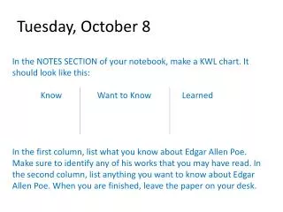
In This Lesson: Unit 2 Microscopes (Lesson 1 of 5). Today is Tuesday, October 8 th , 2013. Pre-Class: Write down three facts you know about microscopes. I will call on each of you for one of them.

E N D
In This Lesson: Unit 2 Microscopes (Lesson 1 of 5) Today is Tuesday,October 8th, 2013 Pre-Class:Write down three facts you know about microscopes. I will call on each of you for one of them. Please get a SMALL paper towel for your pair and take a worksheet from the turn-in box.
Today’s Agenda • Challenge Questions • Unit 2 Pre-Test • History of microscopes • Virtual microscopes • Real microscopes • Painting with fly hair • Where is this in my book? • Academic: P. 169 and following… • Honors: P. 52 and following…
IMPORTANT NOTE • Some of the information we need to learn about microscopes will be discussed when we do the Microscope Lab. • If you’re absent that day, make sure you consult the “Theoretical Data” file and the file called “Lab – Microscope – Information” on my website (Labs section).
History of Microscopes • In the beginning, around 1284, eyeglasses were invented. I still couldn’t see the Bubonic Plague coming. http://www.spectaclesblog.com/wp-content/uploads/2009/09/spectacles-in-the-scriptorium-1352.bmp
History of Microscopes • Around 1590, two Dutch guys (Zacharias Janssen and his son Hans Janssen) put a bunch of lenses together in a tube and noticed that they brought small objects into focus. • Early light microscope! • Janssen also claimed credit for inventing the telescope, but that usually goes to Hans Lipperhey. http://micro.magnet.fsu.edu/optics/timeline/people/antiqueimages/lippershey.jpg http://www.history-of-the-microscope.org/images/Zacharias-Jansen.jpg http://www.microscopy-uk.org.uk/mag/imgjan07/Fig004s.jpg
Light Microscopes • Light microscopes, like the ones we’re using, require light to pass through the sample and into the eyepiece. • Relatively inexpensive. • Can see down to around the level of cell parts – maybe a little smaller. • Can use living things. • Sometimes called optical microscopes.
Then a lot of time passed… • Until we got to the early 1900s. The scanning electron microscope was invented. • Uses a beam of electrons to create images! • It’s big, it’s bulky, it’s expensive, but it’s super-powerful. Does not allow for living specimens. • Samples must sometimes be coated in gold! • Does not require light to pass through an object. • Can view objects about the size of the diameter of an atom.
Types of Microscopes • Scanning Electron Microscope: http://img.directindustry.com/images_di/photo-g/schottky-emission-scanning-electron-microscope-sesem-237074.jpg
Electron Microscope Images http://blog.lib.umn.edu/willow/bioarts/exhibition-ideas/
Electron Microscope Images http://blog.lib.umn.edu/willow/bioarts/exhibition-ideas/
Electron Microscope Images Trichinosis spiralis (microscopic animal parasite from undercooked meat) http://www.photosfan.com/electron-microscope/
Electron Microscope Images http://www.photosfan.com/electron-microscope/
How “Far” Can They See? • Let’s compare. • Scale of the Universe
Scanning Tunneling Microscope • Invented in 1981. • Currently the most powerful microscope available. • Can see three-dimensional objects at the atomic level. • Video!
Willard Wigan • And now for something a bit more…unusual…having to do with microscopes. • This is real.
The Microscope Checklist • So, let’s talk about the parts of a microscope. • Head to your lab tables with your worksheets and notebooks. • Two people from each table should get computers.
Parts of a Microscope 10. Eyepiece Low-Power (Scanning) Objective 7. 9. High-Power Objective 8. Mid-Power Objective 6. Stage Clips Arm 1. 4. 5. Stage Diaphragm/Iris 11. Coarse Focus Knob 3. Light Fine Focus Knob 12. 2. Base
Parts of a Microscope • Arm • Supports the stage, eyepiece, and objectives. • Base • Supports the scope. • Objectives and Eyepiece • Magnify the sample. • Stage and Stage Clips • Hold the slide.
Parts of a Microscope • Coarse Focus • Focuses the image on low power. • Fine Focus • Focuses the image on higher powers. • Diaphragm/Iris • Controls the amount of light entering the objective. • Light • Provides the uh…light.
Meet Your Microscope • Follow this path on your microscope: • When using a light microscope, light comes up from the source through the diaphragm. • It passes through the slide and the object mounted on the slide. • It goes into the objective lens, up the tube and through the eyepiece lens, finally going into your eye.
Handling Your Microscope • First, only handle the scope by the base and arm. • One hand on the arm, the other underneath, as though you’re carrying a giant teacup. • See demonstration! • Give it a shot yourself. Make sure everyone at your table has an opportunity to pick up and set down the scope.
Meet Your Microscope • On low power, the light entering the objective is magnified how many times? • 4x • However, it has to pass through another lens – the eyepiece! • The eyepiece magnifies 10x. • Therefore, to find total magnification, multiply eyepiece magnification by objective magnification. • What are the possible magnifications here? • ALWAYS include magnification with any sketches you draw. It’s like including units with measurements.
Virtual Microscope • Now we’re going to do a little practice on a virtual microscope. • First, log into Quia and complete the Unit 2 Pre-Test as a pair. • When you’re finished, launch the following website: • Virtual Microscope http://www.udel.edu/biology/ketcham/microscope/scope.html • Also found on my website under Supporting Documents. • Close the tour and turn on the checklist. • View the letter “e” on the microscope correctly, then call me over.
Virtual Microscope • http://www.udel.edu/biology/ketcham/microscope/scope.html • Or Supporting Documents – called “Virtual Microscope.” • Close the tour. • Select the slide with the “Letter e” mounted on it. • Select “Checklist” from the upper left corner. • Complete each of the steps. You will need to figure out how to do some of this on your own. • Consider it a scavenger hunt! • When you are finished, show me your results, turn off your computer, and put it away.
Closure • Using a whiteboard, answer the following questions: • Rank the microscopes in terms of increasing magnification power: • Light Microscope, SEM, STM • What is the total magnification on your high-power objective? How did you calculate it? • (show the equation)
Closure • TED: Dee Breger – Visualizing Hidden Worlds Inside Your Body