1 / 2
20 likes | 32 Views
Time-Lapse Imaging Services.<br>https://www.bioimagingtech.com/

E N D
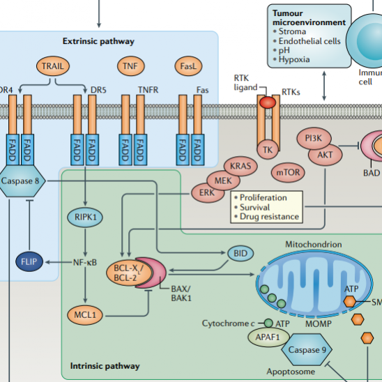
CD BioSciences Launches New Time-Lapse Imaging Services for Scientists CD BioSciences, a US-based biotechnology company focusing on the development of imaging technology, has recently launched its new Time-Lapse Imaging services to assist researchers in a range of fields, including the study of cell migration in chemotaxis assays or wound healing assays, the identification of stem cells and embryos and their development, and the measurement of cell proliferation over time. Time-lapse imaging is a powerful, versatile, and evolving imaging tool for researchers because it can take continuous images at fixed points in time and record the dynamics of what is being observed. Time-lapse imaging of living cells can monitor the response of living cells over time and generate a wealth of image-based information to quantify the behavior of cell populations. Time-lapse imaging has great advantages in the observation and study of cell migration because it is a high-throughput and non-invasive tool for studying cells. CD Biosciences owns advanced imaging equipment and a service support team with extensive experience in the imaging field, and can provide researchers with quality time-lapse imaging services to study cell migration, cell proliferation, etc. CD BioSciences' new time-lapse imaging services can provide high cell viability with a good signal-to-noise ratio. The group advises clients to reduce the exposure time per image and increase the interval between cell recovery images to achieve better results. To minimize phototoxic effects, fluorophores and filters should be matched as closely as possible. Longer wavelength excitation, such as green or red rather than UV or blue, should also be used. Customers should strive to use the highest numerical aperture objective possible for optimal signal detection. Antioxidants may also be added to cell culture medium. Scientists can now explore cellular processes in real-time and acquire insights into intricate biological systems. For example, Time-lapse imaging can be utilized to examine cell migration in chemotaxis tests or to assess wound healing. The movement of cells toward or away from a chemical gradient is known as chemotaxis. In vitro studies of wound healing are typically conducted using wound healing assays. Embryonic and stem cell development can also be studied or identified using time-lapse imaging. Stem cells are critical to regenerative medicine because of their ability to develop into numerous cell types. Embryos go through several stages of development before becoming fully formed organisms, so it is essential to observe them with the new technology. For more information about CD BioSciences' new time-lapse imaging services for scientific research, please visit
https://www.bioimagingtech.com/time-lapse-imaging.html. About CD BioSciences CD BioSciences is a biotechnology company committed to the development of imaging technology for many years. Its scientists can utilize high-content imaging, nanoparticle imaging, imaging flow cytometry, time-lapse imaging, and other techniques to image cell structure, cell migration, cell proliferation, pathogen infection mechanisms, and interactions between protein molecules. In addition, the team is capable of applying powerful analytical software to capture images of cellular structure, subcellular, or tissues to obtain useful information.