Download
1 / 32
320 likes | 594 Views
Emission spectroscopy (mainly fluorescence spectroscopy). Reading : van Holde Chapter 11 Presentation : Nicole Levi: “Probing the interaction between two single molecules: Fluorescence resonance energy transfer between a single donor and a single acceptor” Ha et al. PNAS 93 , 2664

E N D
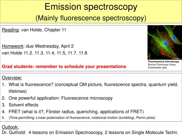
Emission spectroscopy (mainly fluorescence spectroscopy) Reading: van Holde Chapter 11 Presentation: Nicole Levi: “Probing the interaction between two single molecules: Fluorescence resonance energy transfer between a single donor and a single acceptor” Ha et al. PNAS93, 2664 HW: van Holde 11.2. 11.3, 11.4, 11.5, 11.6, 11.7; due Friday, April 8
Quantum mechanics for the purpose of fluorescence 2. singlet Molecules will fluoresce if the emission process has a lifetime that is shorter than the conversion to the triplet state or nonradiative loss of energy. 1. singlet 1. triplet Ground state
Terminology • Luminescence: Process, in which susceptible molecules emit light from electronically excited states created by either a physical (for example, absorption of light), mechanical (friction), or chemical mechanism. • Photoluminescence: Generation of luminescence through excitation of a molecule by ultraviolet or visible light photons. Divided into two categories: fluorescence and phosphorescence, depending upon the electronic configuration of the excited state and the emission pathway. • Fluorescence (emission from singlet state): Some atoms and molecules absorb light at a particular wavelength subsequently emit light of longer wavelength after a brief interval, termed the fluorescence lifetime. Fluorescent molecules are called fluorophores. • Phosphorescence (emission from triplet state): Similar to fluorescence, but with a much longer excited state lifetime.
Example: Fluorescein absorption and emission spectra Stokes shift
Fluorescence instrumentation Excitation monochromator Sample source Emission monochromator Detector Can take absorption and emission spectrum
Fluorescence microscopy • Advantages: • Can label selected features of a sample, eg. Nucleus, DNA, microtubules, specific proteins • Can observe how those molecule behave over time. • Can see (though not resolve) features on nanometer level, even single molecules.
Fluorescence microscopy(here: epi-fluorescence illumination) (See white board)
Normal African Green Monkey Kidney Fibroblast Cells (CV-1) (Olympus web page: http://www.olympusmicro.com) Immunofluorescently labeled with primary anti-tubulin mouse monoclonal antibodies followed by goat anti-mouse Fab fragments conjugated to Rhodamine Red-X. In addition, the specimen was stained with DAPI (targeting DNA in the nucleus).
Fluorescence microscopy (A) and Atomic Force Microscopy images of Oregon-Green-labeled fibrin fibers. Diameters range from 40 to 400 nm.
Fluorescent molecules • Three amino acid have intrinsic fluorescence • Fluorescence of a folded protein is mixture of fluorescence from individual aromatic residues. Most of the emissions are due to excitation of tryptophan. • Tryptophan: • Highest absorptivity and highest quantum strongest fluorescence intensity. • Intensity, quantum yield, and wavelength of maximum fluorescence emission are very solvent dependent. Fluorescence spectrum shifts to shorter wavelength and intensity increases as polarity of the solvent surrounding the tryptophane residue decreases. • Tryptophan fluorescence can be quenched by neighbouring protonated acidic groups such as Asp or Glu. http://dwb.unl.edu/Teacher/NSF/C08/C08Links/pps99.cryst.bbk.ac.uk/projects/gmocz/fluor.htm
Tyrosine • Like tryptophan, has strong absorption bands at 280 nm. • Tyrosine is a weaker emitter than tryptophan, but it may still contribute significantly to protein fluorescence because it usually present in larger numbers. • The fluorescence from tyrosine can be easily quenched by nearby tryptophan residues because of energy transfer effects. • Phenylalanine • Only a benzene ring and a methylene group is weakly fluorescent (product of quantum yield and molar absorbtivity maximum is low. Phenylalanine fluorescence is observed only in the absence of both tyrosine and tryptophane.
http://omlc.ogi.edu/spectra/PhotochemCAD/html/alpha.html Absorption and emission spectra
One Analytical Application • Check for presence of certain proteins, for example, elution from high pressure liquid chromatography. Isolation of melittin, which has one tryptophan residue.
Solvent effects Solvents affect the fluorescence emission spectrum. Two kinds: Specific and general solvent effects. Specific solvent effects: A chemical reaction of the excited state with the solvent. Example: Hydrogen-bonds, acid-base interactions, charge transfer. Changing Fluorescence can be used to detect solvent interactions. 2-anilinonaphthalene fluorescence was changed to hight wavelength by replacing cyclohexan with ethanol. Ethanol forms hydrogen bond.
Solvent effects General solvent effects: Depend on polarizability of solvent increasing dielectric constant shifts fluorescence to higher wavelength. Putting a fluorophore from cyclohexan (low dielectric constant) into water (high dielectric constant), shifts fluorescence to higher wavelengths.
Solvent effects General solvent effect is described by Lippert equation:
Fluorescence Intensity t time Fluorescence decay Absorption N(0) molecules with get excited. Fluorescence intensity is proportional to number of excited molecules. • Decay of excited molecules is a first-order process, with lifetime t. • Decay can happen via three pathways: • Fluorescence with associated intrinsic lifetime to. • Conversion to triplet state (phosphorescence and non-radiative decay). • Non-radiative decay.
Quantum yield When light is absorbed, only a fraction of it is emitted via fluorescence; the rest of the excited molecules decay via other processes. The quantum yield is the ratio of {total number of quanta emitted} to {the total number of quanta absorbed}. The quanta are related to the area under the absorption and emission spectra. t is lifetime of all molecules in excited state, t0 is intrinsic lifetime (lifetime of “fluorescence state”). Corollary: Fluorescence intensity is proportional to product of absorptivity (exctinction coefficient) and quantum yield.
Quantum yield depends very much on environment Changing quantum yield upon binding Increased quantum yield upon binding Application: Staining of DNA in gels. Fluorophores with good DNA binding affinities (often intercalation), extremely large fluorescence enhancements upon binding nucleic acids (some >1000-fold), and negligible fluorescence for the free dyes. Qrel = 1.00 Qrel = 0.46 Qrel = 0.23 SYBR stained dsDNA gel. Excite with UV, emits in visible. (DNA/SYBR Green I complex: Q~0.8; ~300-fold increase over free dye)
Quantum yield depends very much on environment Extinction coefficients were determined for free dye in aqueous solution.
Fluorescence resonance energy transfer (FRET) • When two fluorophores are close together it is possible that one of them absorbs the light (donor), then transfers the energy to the neighboring fluorophore (acceptor), which then emits the light. • The two conditions for this to happen are: • Transition dipole interaction between the two fluorophores (i.e., they need to be close together and aligned. • Significant overlap of the emission spectrum of the donor with the absorption spectrum of the acceptor. Example: Fluorescein (donor) and Alexa-546 (acceptor):
Efficiency of transfer: Fluorescence resonance energy transfer (FRET) Basically, FRET is a great method to determine the distance between two fluorophores (molecules) in the range of ~1-10 nm. Close together FRET signal Far apart (further than Förster radius) no FRET signal • Clever example: Molecular Beacons • used to detect presence of a certain DNA sequence in solution or cells (show on white board).
Fluorescence energy resonance transfer (FRET)Donor-acceptor pairs
Linear polarization of fluorescence • Light to excite fluorophore is now linearly polarized • Emitted fluorescent light will be depolarized Absorption is best for those molecules whose transition dipole is parallel to plane of polarization. • (De-)Polarization of emitted light depends on: • Orientation of emitting transition dipole relative to absorbing transition dipole • Amount of molecular rotation during fluorescent lifetime! • Depolarization of emitted light
Linear polarization of fluorescence Depolarization is described in terms of: • Assume molecules don’t rotate while being excited • depolarization due only to random orientation of molecules with respect to incoming light, q, and angle g: Anisotropy for fluorescence of rhodamine as a function of l of exciting light If there is no molecular rotation, anisotropy will vary between 2/5 (absorbing and emitting trans. dipoles are parallel) and-1/5 (dipoles are perpendicular).
Fluor. anisotropy r r time Linear polarization of fluorescence • Now assume molecules tumble (rotate) before emitting. • depolarization due rotation of molecules. Two extremes: i) molecules don’t rotate before emission r = r0 ii) molecules randomly orient before emitting: r = 0 Time-resolved fluorescence provides a convenient way to measure rotational motion of biological molecules. • … correlation time information about size &shape of molecule • large slow tumbling large molecular weight
Linear polarization of fluorescence Large r slow rotation large molecule Small r faster rotation compact molecule
Perrin plots Instead of pulse illumination, use continuous illumination to measure anisotropy will get average anisotropy ravg. HW 11.6 • … lifetime h … viscosity T … temperature V … volume of molecule
Application of fluorescence to proteins • Analytical detection of presence of proteins • Monitor changes in quantum yield as indication of changing environment (binding, unfolding, etc.) • Effects of energy transfer (FRET). Determine distance of fluorescent groups from each other in 1-10 nm range. • Changes in fluorescence polarization to determine shape and size of molecules (tumbling depends on shape and size) • Monitor (change) in fluorescence parameters to determine stoichiometry, presence of intermediates, binding constants, etc.
Application of fluorescence to DNA • Staining of oligonucleotides in gels • Monitoring the unwinding of double-stranded DNA helicase • Monitoring DNA melting Also: there are tons of reactive fluorophores that can be used to label proteins (Cysteines, primary amines, etc) and DNA. See: Molecular Probes, Inc. http://probes.invitrogen.com/