Download
1 / 22
260 likes | 303 Views
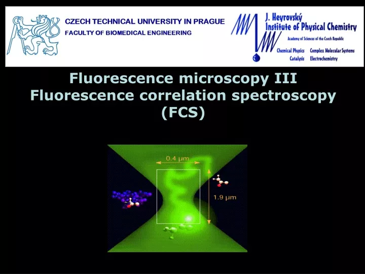
Fluorescence microscopy III Fluorescence correlation spectroscopy (FCS). Detection volume in confocal microscopy:.

E N D
Fluorescence microscopy IIIFluorescence correlation spectroscopy (FCS)
Detection volume in confocal microscopy: the volume from which fluorescence is collected in a confocal (or a multiphoton) is defined by the diffraction limited focusing and the collection efficiency of the objective - Point spread function (PSF) of the microscope typically of femtoliter volume In a diluted solution (~ nM) the average number of molecules in detection volume ~ 1 x ~ 200 nm z ~1 mm The measured fluorescence signal is then very noisy due to fluctuations in the number of molecules in detection volume, their transitions to nonfluorescent states (triplet, …) ~ 3D Gaussian profile
Autocorrelation of fluorescence fluctuations: The timescale of fluorescence fluctuation provides information on the kinetics of the underlying processes. They are studied by correlation analysis. t1 t1 t1 t2 G (t) fluorescence intensity t1/2 t2 t1 t [ms] t [ms] t1/2 – characteristic timescale of the fluctuations Note: Sometimes a different definition of G (t) – converges to 1!!!
Timescale of fluctuations in FCS: The timescale of fluorescence fluctuation provides information on the kinetics of the underlying processes. They are studied by correlation analysis. rotational movement photophysical processes (triplet state, …) G (t) antibunching diffusion t [ms] A single fluorophore molecule emits photons with intervals which are related to its lifetime. More fluorophores in a complex can emit with shorter intervals – investigation of antibunching provides information on molecular oligomerization from Schwille and Haustein: Fluorescence Correlation Spectroscopy
Free diffusion and FCS: The autocorrelation function G (t) is fitted by a theoretical model For free diffusion (e.g. in a solution) and assuming a 3D Gaussian shape of the detection volume following model has been derived: 1/N x direction z direction G (t) tD t [ms] Number of molecules and diffusion time in detection volume wz/w0 – structure parameter, usually ~ 5-8
Free diffusion and FCS: The autocorrelation function G (t) is fitted by a theoretical model When considering the transition to triplet state: characteristic time of triplet transition fraction of molecules in triplet When considering more fluorophore species with different diffusion times: brightness fraction tD1 G (t) tD2 t [ms]
Diffusion coefficient D determination: Diffusion coefficient D of the fluorophore can be calculated from its diffusion time tD The detection volume diameter w0 is usually determined by a calibration measurement with a solution of a fluorophore with known diffusion coefficient for example Rhodamine 6G has D = 426 mm2s-1 In a similar manner concentration can be calculated from N and the detection volume size
E1 DNA compaction investigated by FCS: DNA molecules have pharmaceutical potential in gene therapy, they are however large and negatively charged – difficult transport over cellular membrane Natural solution – compaction of DNA by polycationic molecules such as spermine (+4) amonium/phosphate ratio DNA labelled by intercalating dye PicoGreen is condensed by spermine and the required ration of condenser/base-pair issearched Particle number decreases as the multiple-labelled DNA becomes smaller than the detection volume Adjimatera et al. (2006) Pharm Res 23:1564-1573
Dual-color fluorescence cross-correlation spectroscopy (FCCS): Simultaneous measurement of FCS of 2 different fluorophores excited by 2 different lasers. The emission is divided by an emission dichroic mirror to 2 channels and detected by 2 detectors with appropriate emission filters. Autocorrelation of individual fluorophores and cross-correlation between them can be measured detector emission dichroic major dichroic (double) • Problems: • crosstalk between the two excitation and detection channels • difference in detection volumes in the two channels (diffraction limited focus is larger for longer wavelength)
E2 Dual-color fluorescence cross-correlation spectroscopy (FCCS): Cross-correlation is related to interactions of molecules. Positive cross-correlation indicates that molecules move together (complex). The higher the amplitude of the cross-correlation, the higher complex concentration Negative cross-correlation (anti-correlation) – molecules avoid each other Interaction of 2 membrane proteins: negative control – noninteracting molecules, only crosstalk positive control – double-labeled protein Experiment: Liu et al. (2007) Biophys J 93:684-698
Fluorescence lifetime correlation spectroscopy (FLCS): Uses differences in fluorescence lifetime (instead of in fluorescence spectra) to distinguish contributions to FCS signal Lifetime is sensitive to fluorophore environment FLCS can separate contributions from fluorophores in different environments (different conformation of proteins, …) The method combines FCS with pulsed time-resolved fluorescence spectroscopy (typically TCSPC), arrival time on 2 different scales is measured for each photon photon Laser pulse 2480 1240 3120 Relative Time [ps]
Fluorescence lifetime correlation spectroscopy (FLCS): Each component has its characteristic fluorescence decay (decay pattern) Statistical (numerical) filters (instead of optical filters) are use to separate the photons according to their arrival time after the excitation pulse 5 ns component 2 ns component 5 ns component 2 ns component Filter fj(i) The probability with photons in jth channel contribute to ith pattern. Sum over i equals 1 for each j. For each channel j the measured intensity Ij is a linear combination of patterns:
Fluorescence lifetime correlation spectroscopy (FLCS): Optical filters can improve the data by filtering out scattered light. The statistical filters can do the same – scattered light and noise can be filtered out thanks to their different decay pattern 5 ns component 2 ns component afterpulsing Dark counts (detector afterpulsing) results in a constant background – influence correlation at short lag times (can be misinterpreted as triplet transition) Filter fj(i) Channel j Note: after separating the contributions of individual patterns we can find autocorrelation for each of them and cross-correlations between them If we do not know one of the patterns (it cannot be measured individually), we can still separate the respective contribution by filtering out everything else
E3 DNA compaction investigated by FLCS: What is the compaction mechanism (gradual or all-or-none transition)? For large DNA molecules investigated by single-molecule fluorescence microscopy, but for smaller plasmids below resolution The lifetime of PicoGreen changes upon compaction (change in local polarity) Patterns for uncondensed (4 ns) and fully condensed (3 ns) DNA measured separately and used for investigation of the titration midpoint by FLCS
E3 DNA compaction investigated by FLCS: Patterns for uncondensed (4 ns) and fully condensed (3 ns) DNA measured separately and used for investigation of the titration midpoint by FLCS Good agreement of the autocorrelation of the filtered out components with the pure forms equilibrium between uncondensed and fully condensed form at the midpoint (all-or-none transition) Analysis of cross-correlation between the 2 components reveals further details Humpolíčková et al. (2008) Biophys J94:L17-L19
E3 DNA compaction investigated by FLCS: Analysis of cross-correlation between the 2 components reveals further details Its amplitude between the amplitudes of the two components suggests presence of dynamics between the two forms. Fitting with a model indicates dynamics on ms scale with independent compaction of approximately 5 domains in the DNA molecule. FLCS also showed that another DNA condenser HTAB (+1) exhibits gradual compaction mechanism Humpolíčková et al. (2008) Biophys J94:L17-L19
E4 FLCS and lifetime tuning: Not always is the process we investigate accompanied by a sufficient change in lifetime. Fluorescence lifetime can be influenced externally for example by the vicinity of a conductive surface (quenching) 1.8 ns in supported lipid bilayer (SLB) on ITO surface 5.6 ns in small unilamellar vesicles (SUVs) FLCS G (t) t [ms]
2mm 4nm FCS of planarsamples: In planar samples (lipid bilayers, molecular layers on interfaces, …) the detection volume is reduced to a 2-dimensional Gaussian intensity profile w0≈ 200 nm • Determination of w0 is a problem: • positioning problem: small axial displacement – significant change in w, N and tD. • difference in detection volume in the reference and the sample due to difference in refractive index A need to avoid extrinsic calibration by introducing an intrinsic ruler
Calibration-free FCS: extrinsic calibration avoided by introducing an intrinsic ruler: • axial step between several FCS measurements –Z-scan FCS • parameters of continuous scanning during the FCS measurement – scanning FCS, scanning continuously over a circle of known radius or a line with a defined speed • distance between more points in which FCS is simultaneously measured, multiple measurement points can be generated by: • two overlapping foci generated by doubling the focus by a Wollaston prism (like in DIC) or Michelson interferometer – 2-focus FCS. • different pixels of a microscope image (recorded for example in TIRF configuration) – Image correlation spectroscopy (ICS, STICS, …) • combination of scanning and imaging in laser scanning microscopy (LSM) – known pixel size and scanning speed – RICS, STICS, …
DZ Z-scan FCS: known axial step between measurements serves as intrinsic calibration parabolic dependence of w2, N and tD on DZ: Determination of surface concentration cS and diffusion coefficient D of the fluorophore
ICS: spatial correlation between image pixels (distances along x and y axes play the role of lag time) The amplitude of the correlation peak is inversely proportional to fluorophore density • additional temporal information allows investigation of diffusion: • correlations between images in a temporal series (spatio-temporal ICS – STICS) diffusion – broadening of the peak oriented flow – broadening + shift • possibility to construct velocity maps • temporal resolution defined by imaging speed t • imaging by LSM with defined scanning speed spatial correlation contains temporal information (Raster image correlation spectroscopy – RICS), 2 axes – 2 timescales Hebert et al. (2005) Biophys J 88:3601-3614
Acknowledgement The course was inspired by courses of: Prof. David M. Jameson, Ph.D. Prof. RNDr. Jaromír Plášek, Csc. Prof. William Reusch Financial support from the grant: FRVŠ 33/119970