Download

1 / 19
200 likes | 377 Views
Review for Cells. History of Microscopy. Hooke observes first cells (cork) -1665 Leeuwenhoek – animalcules (living cells)- 1674 Jump to late 1830s Schleiden – plants made of cells 1838 Schwann – animals made of cells 1839 Brown - nucleus Virchow – Cells make more cells – 1850s

E N D
History of Microscopy • Hooke observes first cells (cork) -1665 • Leeuwenhoek – animalcules (living cells)- 1674 • Jump to late 1830s • Schleiden – plants made of cells 1838 • Schwann – animals made of cells 1839 • Brown - nucleus • Virchow – Cells make more cells – 1850s • RESULT: THE CELL THEORY
Cell Theory • All living things made up of one or more cells • Cells are the basic unit of structure and function in all living things (no smaller LIVING thing than a cell) • Cells come from previous cells
Types of microscopes • Light microscopes • Stereo • Low magnification, 3D, color, surface structures, light, often used to compare two things (like bullets…(ballistics) • Compound • Magnifies up to 2000, 2D, color, cross sections, light, two lenses • Electron Microscopes • Scanning • Magnifies up to 200,000, 3D, surface, electrons, B and W. Shows surface structures at high resolution • Transmission • Magnifies up to 2,000,000, 2D, cross section, electrons, BW, shows internal cell structures (organelles at high resolution)
Compound microscope • Inverts and flips the image • Move slide right, appears to move left • Move slide up, appears to move down • High power smaller field, less light getting in, so typically need to open up the diaphragm • Center so that high power (smaller field) will pick up what you are “viewing” • Magnification increases size, resolution discriminates between two points that are very close together.
Cell Diversity • Different shapes and sizes due to different functions • Form fits function • SA/V ratio. Cells can’t get too big or they will die. When the surface area can’t feed the volume, the cell divides
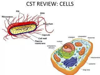
Cell Types • Prokaryotes • Only single cells • Very small • No membrane bound organelles • Ribosomes, membrane, cell wall, cytoplasm, nucleic acids • Three major shapes: bacillus, spirillium, coccus
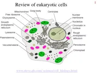
Cell Types • Eukaryotes • One t0 billions of cells • Simple to very complex • All contain a nucleus (one or more) • All kingdoms except Prokaryote • Plants have chloroplasts, cell walls, large central vacuole • Animals have small vacuoles, centrioles, no cell wall • 10-100x bigger than prokaryotes
Cell Organelles (see pictures • Look at diagrams provided and the six “processes”. Trace the process creating a flow chart that covers key points. • Make a protein • Endocytosis and exocytosis • Energy • Cell division • Movement of cells • Molecules, ions and types of transport
Cell Transport • Phospholipids, Proteins, carbohydrates, cholesterol • Nucleus, cytoplasm, ER, Golgi • Cholesterol: flexibility • Carbohydrates: identification tags
Membrane Function • CO2, H2O, O2, small non-polar compounds , N2 • Small, non-polar • Membrane proteins – very specific for particular molecules • Amino acids, glucose, ions • Charged, larger • Energy is needed when molecules or ions need to move AGAINST the gradient (from low to high)
Membrane Function • No energy needed when flowing with the gradient (high to low) • Fluid mosaic: fluid do to movement of molecules through the lipid bilayer, mosaic due to different molecules that make up the membrane. • Semi-permeable, allows some molecules, but not others, doesn’t discriminate between “good” and “bad”
Transport through the membrane • Passive transport • Diffusion – membrane NOT required (food coloring, perfume) • Facilitated Diffusion (protein mediate) • Osmosis • Active transport • Protein mediated • Vesicle mediate
Osmosis and Tonicity • Osmosis is the movement of water only. • In our examples, when water moves, the solute does not. • Tonicity refers to the concentration of SOLUTE (as compared to another location) • Hypertonic (high solute, low water) • Hypotonic (low solute, high water) • Isotonic ( equal amounts of solute) • Water moves from hypotonic to hypertonic (low solute high solute, or HIGH water, to LOW water
Living cells in different tonicities • What is the situation in which cells crenate • What type of cells do this? • What type of cells undergo cytolysis? Under what conditions • Why is high turgor good for plants? • When can plasmolysis occur, when a solute isn’t present?
Active Transport vs. Vesicle Transport • Number of molecules moving at one time dictates active (few molecules) vs vesicle (many molecules) • Both require ATP • Active transport is through a protein, against the gradient (low to high) • Vesicle transport wraps a membrane around the molecules, creating a “cab”. doesn’t require a gradient
Vesicle transport • Vesicles move many molecules at a time either out of the cell or into the cell • Exocytosis –OUT of the cell • Example: hormones, mucus, cell waste • Endocytosis - into the cell (see next page) • Phagocytosis – larger particles (proteins, bacteria, etc) • Pinocytosis – dissolved particles (liquid) • Hormones are released through exocytosis (too many molecules • Phagocytosis – WBC eat bacteria!