Download
1 / 163
1.63k likes | 1.87k Views
CNS & PNS. Outline. CNS Basic CNS Pattern Spinal Cord Anatomy Reflexes Trauma Meninges & CSF Blood Brain Barrier Brain. PNS ANS Sympathetic Parasympathetic Neurotransmitters Somatic Cranial Nerves Spinal Nerves. Basic Pattern of the Central Nervous System. Figure 12.5.

E N D
Outline • CNS • Basic CNS Pattern • Spinal Cord • Anatomy • Reflexes • Trauma • Meninges & CSF • Blood Brain Barrier • Brain • PNS • ANS • Sympathetic • Parasympathetic • Neurotransmitters • Somatic • Cranial Nerves • Spinal Nerves
Basic Pattern of the Central Nervous System Figure 12.5
Spinal Cord • Functional Areas • Dorsal Root • Ventral Root • Spinal Tracts • Ascending Tracts • Dorsal column tract • Anterolateral System • Descending Tracts • Corticospinal Tract
Reflexes • A reflex is a rapid, predictable motor response to a stimulus • Reflexes may: • Be inborn or learned (acquired) • Involve only peripheral nerves and the spinal cord • Involve higher brain centers as well
Reflex Arc • There are five components of a reflex arc • Receptor • Sensory neuron • Integration center • Motor neuron • Effector
Reflex Arc Figure 13.12
Reflex Classification • Classified Functionally • Somatic Reflexes • Autonomic reflexes
Accommodation reflex Achilles reflex Biceps stretch reflex Brachioradialis reflex Corneal reflex (also known as the blink reflex) Crossed extensor reflex Gag reflex Mammalian diving reflex Patellar reflex (knee-jerk reflex) Photic sneeze reflex Plantar reflex (Babinski reflex) Pupillary reflex Quadriceps reflex Salivation Scratch reflex Sneeze Tendon reflex Triceps stretch reflex Vestibulo-ocular reflex Withdrawal reflex Yawn Reflexes in Adult Humans
Flexor and Crossed Extensor Reflexes • The flexor reflex is initiated by a painful stimulus (actual or perceived) that causes automatic withdrawal of the threatened body part • The crossed extensor reflex has two parts • The stimulated side is withdrawn • The contralateral side is extended
Crossed Extensor Reflex Figure 13.17
Mammalian Diving Reflex • Submerging the face into water causes the mammalian diving reflex • Includes three factors: • Bradycardia • Peripheral vasoconstriction • Blood shift • When the face is submerged, receptors that are sensitive to water within the nasal cavity and other areas of the face supplied by cranial nerve V (trigeminal) relays the information to the brain and then innervates cranial nerve X (vagus).
Photic Sneeze Reflex • A medical condition by which people exposed to bright light sneeze. • Occurs in 17% to 25% of humans • The probable cause is a congenital malfunction in nerve signals in the trigeminal nerve nucleus. • Overstimulation of the optic nerve triggers the trigeminal nerve, and this causes the photic sneeze reflex.
Yawn • A reflex of deep inhalation and exhalation associated with being tired, with a need to sleep, or from lack of stimulation. Pandiculation • It is claimed to help increase the state of alertness of a person. It could possibly be from lack of oxygen. • The exact causes of yawning are still unknown.
Spinal Cord Trauma: Paralysis • Paralysis – loss of motor function • Flaccid paralysis – severe damage to the ventral root or anterior horn cells • Spastic paralysis – only upper motor neurons of the primary motor cortex are damaged
Spinal Cord Trauma: Transection • Cross sectioning of the spinal cord at any level results in total motor and sensory loss in regions inferior to the cut • Paraplegia – transection between T1 and L1 • Quadriplegia – transection in the cervical region
Poliomyelitis • Destruction of the anterior horn motor neurons by the poliovirus • Early symptoms – fever, headache, muscle pain and weakness, and loss of somatic reflexes
Amyotrophic Lateral Sclerosis (ALS) • Lou Gehrig’s disease • Symptoms – loss of the ability to speak, swallow, and breathe
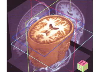
The Brain • Composed of wrinkled, pinkish gray tissue • Surface anatomy includes cerebral hemispheres, cerebellum, and brain stem
Adult Neural Canal Regions Figure 12.3a, b
Adult Neural Canal Regions Figure 12.3c
Adult Neural Canal Regions Figure 12.3c, e
Ventricles of the Brain Figure 12.6
Protection of the Brain • The brain is protected by bone, meninges, and cerebrospinal fluid • Harmful substances are shielded from the brain by the blood-brain barrier
Meninges • Three connective tissue membranes that lie external to the CNS – dura mater, arachnoid mater, and pia mater • Functions of the meninges include: • Cover • Protect • Contain • Form
Meninges Figure 12.20a
Dura Mater • Leathery, strong meninx composed of two fibrous connective tissue layers • The two layers separate in certain areas and form dural sinuses • Three dural septa extend inward and limit excessive movement of the brain • Falx cerebri • Falx cerebelli • Tentorium cerebelli
Dura Mater Figure 12.21
Arachnoid Mater • The middle meninx, which forms a loose brain covering • It is separated from the dura mater by the subdural space
Arachnoid Mater Figure 12.20a
Pia Mater • Deep meninx composed of delicate connective tissue that clings tightly to the brain
Cerebrospinal Fluid (CSF) • Watery solution similar in composition to blood plasma • Contains less protein and different ion concentrations than plasma • Forms a liquid cushion that gives buoyancy to the CNS organs
Cerebrospinal Fluid (CSF) • Circulates through the ventricle system of the brain to the brainstem • Aided by circulatory, respiratory and postural changes • Barrier between the blood in the capillaries of the choroids plexus and the
Choroid Plexuses • Clusters of capillaries that form tissue fluid filters, which hang from the roof of each ventricle • Have ion pumps that allow them to alter ion concentrations of the CSF • Help cleanse CSF by removing wastes
Choroid Plexuses Figure 12.22a
Blood-Brain Barrier • Protective mechanism that helps maintain a stable environment for the brain • Bloodborne substances are separated from neurons by: • Continuous endothelium of capillary walls • Relatively thick basal lamina • Bulbous feet of astrocytes
Blood-Brain Barrier: Functions • Selective barrier • Absent in some areas • Responds to stress
Blood-Brain Barrier: Functions • Reduce the immune systems access to the brain • Comprised of the cells that make up the smallest blood vessels of the brain • Anatomical structures • Physiological transport systems • Accounts for some drug actions • Morphine vs. heroin • Many substances are not lipid soluble • Glucose and other important brain substrates
Brain • Brainstem • Midbrain • Pons • Medulla Oblongata • Cerebellum • Forebrain • Diencephalon • Cerebrum • Limbic System
Brain Stem Figure 12.15
Midbrain • Located between the diencephalon and the pons • Midbrain structures include: • Cerebral peduncles • Cerebral aqueduct • Various nuclei
Midbrain Nuclei Figure 12.16a
Midbrain Nuclei • Nuclei that control cranial nerves III (oculomotor) and IV (trochlear) • Corpora quadrigemina • Superior colliculi • Inferior colliculi • Substantia nigra • Red nucleus – largest nucleus of the reticular formation
Pons Figure 12.6b
Pons • Bulging brainstem region between the midbrain and the medulla oblongata • Forms part of the anterior wall of the fourth ventricle • Fibers of the pons: • Connect higher brain centers and the spinal cord • Relay impulses between the motor cortex and the cerebellum • Origin of cranial nerves V (trigeminal), VI (abducens), and VII (facial) • Contains nuclei of the reticular formation
Reticular Formation Figure 12.19
Reticular Formation: RAS and Motor Function • RAS – reticular activating system • Sends impulses to the cerebral cortex to keep it conscious and alert • Filters out repetitive and weak stimuli • Motor function • Helps control coarse motor movements • Autonomic centers regulate visceral motor functions