Download
1 / 22
220 likes | 360 Views
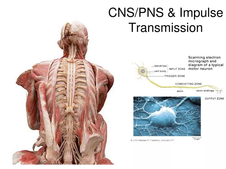
CNS/PNS & Impulse Transmission. Autonomic NS- “subconscious” Regulate activity of smooth muscle (digestive), cardiac muscle (heart) & certain glands (midbrain) Structures involved –neurons – integration within the brain • Receives input, ready for action Somatic NS- “conscious”

E N D
Autonomic NS- “subconscious” Regulate activity of smooth muscle (digestive), cardiac muscle (heart) & certain glands (midbrain) Structures involved –neurons – integration within the brain • Receives input, ready for action Somatic NS- “conscious” Means consciously perceived sensations – excite skeletal muscle to move muscles/skeleton Structures involved - neuron connects CNS to organ or muscle
Reaction in Action: Click Above
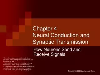
So here’s how it ALL goes down!(fast and ugly….but the whole ball of wax!) • Sensory neuron stimulated • Neuron becomes positive; K channels open • Neurotransmitters released into synapse space • Push/excite all nearby neurons Watch this at work: McGraw-Hill: K/Na pump and synaptic cleft movement animations
What happens when there is a ‘gap’ in impulse transmission? What does a severed spinal cord mean here?
PNS: HUMAN VISION • Binocular Vision • Depth perception • Photo-sensetiveCells • Rods: light/dark • Cones: color • Optic nerve: wired directly from rods and cones to eye through optic chiasmata
MusclePrimary Function Medial rectus moves eye towards nose Lateral rectus moves eye away from nose Superior rectus raises eye Inferior rectus lowers eye Superior oblique rotates eye Inferior oblique rotates eye
Eye Structures Cornea Tough transparent outer layer Light enters Fluid chamber behind (AH) Iris Disk of colored contractile tissue Autonomously contolled Pupil (Regulated by iris) determines amount of VLS entering the eye Attached to tiny muscles to lens and eye Lens Flexible and transparent piece of protein Iris (Small muscles) attached Convex: flips images over (corrected in visual cortex of brain)
Vitreous humor Jelly-like fluid Transparent Shape Aqueous humor Lubricates lens (after cornes) Retina Contrast Cones/Rods Allows you to see color…sometimes??? Optic nerve Sends messages to brain Sclera ‘whites’ of the eyes ** Tapetum Reflective membrane; night vision
Magic….or • Optical illusions- overstimulation of photosensitive cells in retina results in confusion, dizziness and in some cases even nausea and seizure Enjoy the illusions!
Useful Resources: • Cow eye dissection video • Cow eye dissection worksheet • Cow eye dissection lab (includes worksheets) • Cow eye dissection quiz
TODAY’S TASK: Reflex lab • Today, with a partner, you will determine your reflex ‘arc’: from sensory input to brain processing to motor reaction • First trial = silence • Second trial = distraction