Download
1 / 18
180 likes | 310 Views
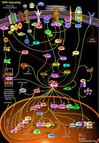
Expression of an antioxidative enzyme in the normal and pakinsonian brain presented by Simone Abercrombie Bio 475. van Muiswinkel F.L., R. A. I. de Vos, J.M. Bol, G. Andringa, E.H. Jansen Steur, D. Ross, D. Siegel and B. Drukarch. Signaling. Neuron. Dopaminergic terminal.

E N D
Expression of an antioxidative enzyme in the normal and pakinsonian brainpresented bySimone AbercrombieBio 475 van Muiswinkel F.L., R. A. I. de Vos, J.M. Bol, G. Andringa, E.H. Jansen Steur, D. Ross, D. Siegel and B. Drukarch
Dopaminergic terminal Prototypic dopaminergic terminal with cycle of synthesis, storage, release and removal of dopamine. (Cooper, Bloom & Roth, 1996)
Parkinson’s disease Dopamine The basal ganglia is responsible for motor function. The cells in this area need a proper balance of dopamine, a neurotransmitter. In Parkinson's, cells that produce dopamine begin to degenerate. Loss of dopamine causes the nerve cells of the striatum to fire out of control, leaving patients unable to direct or control their movements in a normal manner. Studies have shown that Parkinson's patients have a loss of 80 percent or more of dopamine-producing cells in the substantia nigra.
Major neural pathways in normal and Parkinsonian basal ganglia, (Vermeulen, 1994) . The thickness of the arrows represents the strength of the signal.
Why do cells degenerate? 1. Free radicals • Free radicals are highly unstable chemicals damage cell structures. Most stable compounds in the body possess a pair of electrons. When an electron of the electron pair gets stripped away its called a free radical. They are highly unstable because the seek other compounds and break bonds between stable compounds causing a chain reaction. • Free radicals can come from environmental pollution, radiation, cigarette smoke, chemicals, and herbicides.
Why do cells degenerate? 2. Genetics There are several genetic mutations and a few genes that are linked to PD. Inheriting mutated genes may me the same genes that are altered sporadically by environmental factors and toxins. • alpha-synuclein-This gene was mutated in families with familial PD • Parkin-encoded into a protein which functions to help cells break down and recycle proteins • DJ-1-normally helps control gene activity and protect cells from oxidative stress • PINK1-when it is mutated it increases vulnerability to cellular stress • LRRK2-linked to late onset of familial PD and a small percentage in sporadic PD
Why do cells degenerate?3. Aging • In some individuals, the normal, age-related wearing away of dopamine-producing neurons accelerates. This theory is supported by the fact that the loss of antioxidative protective mechanisms is associated with both Parkinson's disease and increasing age.
NAD(P)H:Quinone oxidoreductase (NQO1) • detoxication enzyme • catalyses the two-electron reduction of DAQs into DAhydroquinone, a relatively redox stable entity that not only lacks major electrophilic reactivity but is also amenable to further detoxication by phase II enzymes
Rationale • examine the cellular expression of NQO1 in the brain of a large series of idiopathic(no known cause) PD patients and age-matched controls • data on the cellular localization of NQO1 in the Parkinsonian SNpc is lacking • investigate the potential role of NQO1 in the pathogenesis of PD
Specificity of NQO1 immunocytochemistry • Immunohistochemical staining with antibodies raised against NQO1 protein shows expression of NQO1 in normal respiratory epithelium and NSCLC (adenocarcinoma). • No expression in small cell lung cancer (SCLC), surrounding lymphoid cells or supporting stroma. A) SCLC (asterisk) tissue with normal respiratory epithelium (brown); (B and C) non-NSCLC (double asterisks) tissue with scattered coal deposits and normal respiratory epithelium (C, brown); (D) NSCLC tissue immunostained for NQO1 with AEC.
Expression of NQO1 in the control substantia nigra, pars compacta (SNpc) • NQO1 immunoreactivity could be detected in three major cell types: melanized dopaminergic neurons, astrocytes, and vascular endothelium. • NQO1 staining was not detected when antibodies were omitted or replaced by C100 control hybridoma supernatant (data not shown). • cytoplasmatic staining of NQO1 was observed in neuromelanin containing dopaminergic neurons • A, asterisks points out the neuromelanin pigment • B, astroglial cells • C, vascular endothelium • D shows a NQO1-immunopositive astrocyte in a Parkinsoninan SNpc.
…contd • Expression of NQO1 and glial markers in the normal SNpc. • Arrows in A and B indicate (NQO1 immunopositive) astrocytes.
Expression of NQO1 in the Parkinsonian substantia nigra, pars compacta • B–D show high-power magnifications of subfields of the neuromelanin containing neurons shown in panel A. • Reactive astrocytes are indicated by arrows • Arrowheads denote NQO1-immunonegative intracytoplasmatic Lewy bodies. • Reactive astrogliosis and a large number of NQO1 immunopositive dopaminergic neurons in panels C and D.
Patients with intermediate stage PD(A–C) • End-stage PD (D-F). • Intermediate-stage PD is characterized by the presence of marked gliosis and intense NQO1-immunostaining of astroglial cells (arrows) • End-stage PD NQO1 expression is limited to vascular endothelial cells (double arrow). …contd
Discussion • The degenerative process in the PD SNpc is accompanied by an increase in neuronal and astroglial NQO1 expression • NQO1 is expressed either as fibrous astrocytes, pigmented dopamine neurons, or a combination of both. • The increase in NQO1 expression in the Parkinsonian SNpc indicates a possible correlation between the expression of NQO1 and ongoing degeneration of dopaminergic neurons. • Expression of NQO1 increases when degeneration of neurons is actively taking place. PD patient’s condition is characterized into three different stages: early, intermediate, and end. At the end stage most of the neurons have been depleted (depletion inactively taking place) and NQO1 immunoreactively is nearly not present.
References References: • Beyer R.E, J. Segura-Aguilar, S. Di Bernado, M. Cavazzoni, R. Fato, and D. Fiorentini. 1997. The two-electron quinone reductase DT-diaphorase generates and maintains the antioxidant (reduced) form of coenzyme Q in membranes. Molecular aspects of medicine. 18:15–23. • Cadenas E. 1995. Antioxidant and prooxidant functions of DT-diaphorase in quinone metabolism. Biochemical Pharmacology. 49:127–140. • Drukarch B. and F.L. van Muiswinkel. 2000. Drug treatment of Parkinson’s disease: time for phase II. Biochemical Pharmacology. 59:1023–1031. • Drukarch B. and F.L. van Muiswinkel. 2001. Neuroprotection for Parkinsons’s disease: a new approach for a new millennium. Expert opinion on investigational drugs. 10:1855–1868. • Harada S., C. Fujii, A. Hayashi and N. Ohkoshi. 2001. An association between idiopathic Parkinson’s disease and polymorphisms of phase II detoxification enzymes: glutathione S-transferase M1 and quinone oxidoreductase 1 and 2. Biochemical and biophysical research communications. 288:887–892. • Segura-Aguilar J. and C. Lind. 1989. On the mechanism of the Mn3+-induced neurotoxicity of dopamine: prevention of quinone-derived oxygen toxicity by DT diaphorase and superoxide dismutase. Chemico-biological interactions. 72:309–324. • van Muiswinkel F.L., R. I. de Vos, J.M. Bol, G. Andringa, E.H. Jansen-Steur, D. Ross, D. Siegel and B. Drukarch. 2003. Expression of NAD(P)H:quinone oxidoreductase in the normal and Parkinsonian substantia nigra. Neurobiology of Aging. 25:1253-1262.