Download
1 / 26
360 likes | 568 Views
Cell Signaling. Lecture 27 Chapter 16. Eyeless. Cells can exist as single celled organisms or be part of a multi-cellular organism How do they know what is happening around them? Cells do not have ‘eyes’, ‘noses’, or ‘ears’

E N D
Cell Signaling Lecture 27 Chapter 16
Eyeless • Cells can exist as single celled organisms or be part of a multi-cellular organism • How do they know what is happening around them? • Cells do not have ‘eyes’, ‘noses’, or ‘ears’ • Answer: Cells communicate with others and have mechanisms to sense their environments using a variety of methods as we will uncover - whichever method is used it involves a very important principle called SIGNAL TRANSDUCTION
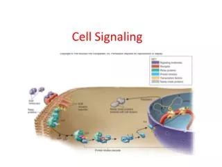
Signal Transduction 16_02_Signal_transduct.jpg The conversion of a signal, of some type, from one physical form to another. Electrical impulses are converted to sound waves that we hear Signal molecules are received by target cells via receptors and converted to other intracellular forms
Cell Signaling types • Endocrine - hormones, long distance • Paracrine - local vicinity • Neuronal - very short distances • Contact-dependent - physical contact • Autocrine - act on self
The same signal molecule may interact with different cells with entirely different effects. Here is an example of acetylcholine 16_05_target_cells.jpg Acetylcholine has a role in both branches of your nervous system It has a half-life of about 2 minutes.
Cells generally respond to a combination of signals. The same cell may have different outcomes to various signal combinations 16_06_extracellular_sig.jpg Cells that DO NOT RECEIVE SIGNALS DIE The default pathway=
How do these signals work At which level? Where? How many?
General Plan of Action 16_07_change_behavior.jpg
Each cell responds to a limited set of signals - why? • These signals change the activity of internal cellular proteins which chances the behavior of the cell • These signals follow a chain of events known as the signal cascade • A system of relayinginformation from the site of reception to the point of action • Normally the signal is amplified too - a small input is quickly converted to a large response
General Overview 2 16_08_cascades.jpg
Some signal molecules act at the cell surface whilst others can enter the cell readily and act inside such as steroids. 16_09_molecules_bind.jpg The red signal molecule has a target receptor to which it binds and that’s that. Note that it is usually hydrophilic. Other receptors enter the cell. If they must pass through the membrane without P’s they are..
A simple example of how a steroid works. -The signal can enter the cell through the membrane and bind to its target protein. -This is now able to enter through the nuclear pore and control transcription directly of certain target genes 16_12_cortisol.jpg
There are three classes of cell surface receptor: ion-channel linked G-protein linked Enzyme linked 16_14_3_basic_classes.jpg
All nerve impulses are generated via ion-channel linked receptors • The release of neurotransmitter causes the ion-channel on the target neuron to allow the passage of ions (which?) into the cell. This action is propagated through the nerve cell along its axon.
Many interactions taking place within the cell act to turn on or off proteins. These are known as molecular switches. 16_15_molec_switches.jpg
G-protein linked receptors • These form the largest class of cell-surface receptors so far identified - with many hundreds known • They have a common structure • A single polypeptide that passes 7 times back and forth across the plasma membrane • These receptors can bind to all sorts of signal molecules - proteins, small peptides, amino acids, even fatty acids • When bound by its extracellular signal molecule the receptor undergoes a physical change, which permits it to activate a G-protein located in the inside surface of the plams membrane
The key to the regulation of the cell is the behavior of the G-protein. It is a trimer of three subunits - When the three units are associated with each other there is no activity When the receptor is activated it causes the disassociation of the subunits by replacing the bound GDP with GTP. Each of the two subunits is active to catalyze other cellular molecules. The GTP is eventually converted to GDP and the 3 subunits reunite. 16_17_Gprot_dissociate.jpg
Continued from previous - details on how the subunit is inactivated 16_18_Gprot_subunit.jpg
Some G-proteins activate ion channels. Here we see a schematic showing how the K pump in the heart is controlled 16_19_open_K_chan.jpg
Some G proteins activate enzymes directly 16_20_second_messeng.jpg
Some cellular responses are quick, whilst others are slow. 16_23_slowly_rapidly.jpg
Calcium has a very important role to play as an intercellular messenger. As we know the concentration of calcium is extremely low in the cytoplasm of a typical cell, compared to the outside and to that of the ER. In this example fertilization results in the wave of receptors opening up to permit the influx of calcium into the cell. This results in a change in the cell surface which both initiates cell division and prevents other sperm from entering the cell. 16_26_Fertilization.jpg
The speed at which signaling cascades operate is clearly illustrated by the photoreceptors of the eye. The human eye has two forms of receptors - rods and cones The cones are further divided depending on the wavelength of light they respond to - red, green, and blue 16_28_photoreceptor.jpg
The more complex receptors are illustrated by tyrosine kinases. These act in trans on each other to phosphorylate certain regions. The phosphorylated regions bind other factors in the cell. 16_30_rec_tyro_kinase.jpg