Download
1 / 24
290 likes | 627 Views
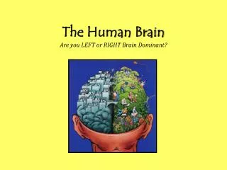
E5 The human brain. 489 - 498. Assessment Statements. E.5.1 Label, on a diagram of the brain, the medulla oblongata, cerebellum, hypothalamus, pituitary gland and cerebral hemispheres. E.5.2 Outline the functions of each of the parts of the brain listed in E.5.1.

E N D
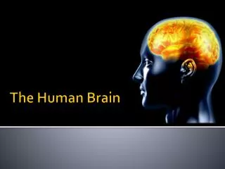
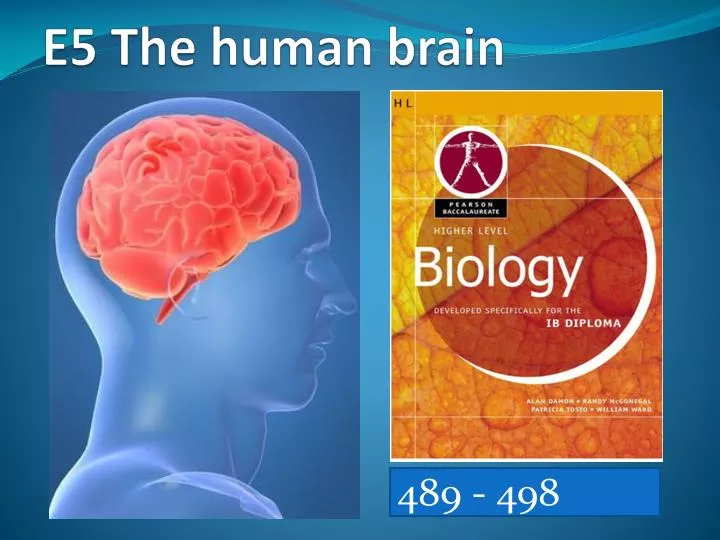
E5 The human brain 489 - 498
Assessment Statements • E.5.1 Label, on a diagram of the brain, the medulla oblongata, cerebellum, hypothalamus, pituitary gland and cerebral hemispheres. • E.5.2 Outline the functions of each of the parts of the brain listed in E.5.1. • E.5.3 Explain how animal experiments, lesions and fMRI (functional magnetic resonance imaging) scanning can be used in the identification of the brain part involved in specific functions. • E.5.4 Explain sympathetic and parasympathetic control of the heart rate, movements of the iris and flow of blood to the gut. • E.5.5 Explain the pupil reflex. • E.5.6 Discuss the concept of brain death and the use of the pupil reflex in testing for this. • E.5.7 Outline how pain is perceived and how endorphins can act as painkillers.
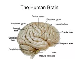
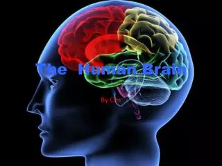
On the diagram of the brain, label the following: medulla oblongata; cerebellum; hypothalamus; pituitary gland & cerebral hemispheres cerebral hemisphere hypothalamus cerebellum pituitary gland medulla oblongata spinal cord
Functions of each of the parts of the brain Part of the brain Function(s) medulla oblongata controls autonomic functions of the body such as: heart rate; blood pressure; ventilation; swallowing; vomiting; digestion & cranial reflexes Cerebellum coordinates unconscious functions, such as movement and balance Hypothalamus links nervous and endocrine systems; produces hormones secreted by posterior pituitary; controls hormonal secretion by pituitary; maintains homeostasis such as; control of body temperature, hunger, thirst, fatigue, circadian cycles pituitary gland the posterior lobe stores and releases hormones produced by the hypothalamus and the anterior lobe, and produces and secretes hormones regulating many body functions cerebral hemispheres act as the integrating centre for high complex functions such as learning, memory and emotions
Use of animal experiments to identify the brain part involved in specific functions • experiments involves surgery to remove part of the scull to access the brain • animal must be alive during this procedure • different regions of the brain are stimulated and the response of the animal observed • primates were often used but it raised ethical issues due to their genetic similarities to humans & the pain and suffering the procedure caused • example :- Pierre in 1820s showed that removing thin slice of tissue from the cerebellum of rabbits & birds resulted in animals displaying lack of muscular coordination & poor balance with no other obvious effects
Lesions & their uses in identification of the brain parts involved in specific functions • lesionsare any abnormalities in the brain tissue of an organism • lesions could be due to damage to the brain tissue as a result of accidents, stroke, tumour or deliberate injury • lesions indicates effect of loss of a brain tissue by comparing an organism to a normal one • e.g. split brain patients led to understanding different functional roles of left and right hemispheres of the brain • many actions of the body involve different areas of the brain • damage may be to many parts of the brain, thus making it difficult to interpret due to complexity of reactions
fMRI (functional magnetic resonance imaging) scanning • fMRI stands for functional magnetic resonance imaging • fMRI records changes in blood flow to the brain • active parts of the brain have increased blood flow • but not all brain activity is detected by MRI • a subject is given a stimulus which is designed to stimulate brain activity • MRI links stimulus with certain part of the brain • brain activity visualized by coloured images
Use of fMRI scanning in identification of the brain parts involved in specific functions • fMRI gives a more specific knowledge of stimulated area of the brain • e.g. it is used to study (diagnose) ADHD, dyslexia, recovery from strokes, music comprehension etc. • fMRI is non-invasive (no damage to brain), can study healthy subjects • involves recording increased blood flow & supply of oxygen to the active parts of the brain • good spatial but poor temporal resolution • it requires interpretationof coloured images • there could be a problem of statistical interpretations of models
CENTRAL NERVOUS SYSTEM (CNS) PERIPHERALNERVOUS SYSTEM (PNS) BRAIN AND SPINAL CORD PERIPHERAL NS SOMATIC NERVOUS SYSTEM (voluntary) AUTONOMIC NERVOUS SYSTEM (involuntary) MOTOR NEURONES TO INTERNAL ORGANS SENSORY AND MOTOR NEURONES TO / FROM SKELETAL MUSCLE SYMPATHETIC NERVOUS SYSTEM (involuntary) PARASYMPATHETIC NERVOUS SYSTEM (involuntary) CONTROLS ORGANS WHEN BODY IS AT REST CONTROLS ORGANS IN TIMES OR STRESS The Organisation of the Nervous System NERVOUS SYSTEM
Series of ganglia The sympathetic nervous system (SNS) The cell bodies of its motor neurones lie in ganglia outside the spinal cord PUPILS SALIVARY GLANDS It prepares the body for action HEART The transmittersecreted at these synapses is usually nor adrenaline – which stimulates organ activity BRONCHI LIVER STOMACH/ SMALL INTESTINE The heart beats faster, eyes get wider i.e. the pupil dilates to improve vision, “sinking” feeling in the stomach due to decreased blood supply to the gut. ADRENAL GLAND / KIDNEYS LARGE INTESTINE BLADDER / GENITALS SNS functions are FIGHT OR FLIGHT
The Parasympathetic Nervous System (PNS) All nerve pathways begin in the brain, or at the top or bottom of the spinal cord EYE SALIVARY GLANDS The neurones keep going ‘till right inside the organ. Here they synapse with a motor neurone BRONCHI HEART The transmittersecreted at these synapses is acetylcholine and this has an inhibitory effect on the organ STOMACH PYLORIC SPHINCTER PANCREAS Heart rate decreases, increased blood flow to the gut, constricts pupil to protect retina LARGE INTESTINE, ANAL SPHINCTER BLADDER PNS functions are REST AND DIGEST GENITALS
Effects of Sympathetic & Parasympathetic NS Sympathetic NS Parasympathetic NS • secretes noradrenaline or norepinephrine • accelerates heart rate • causes widening (dilation) of the pupils • in gut, stomach, pancreas, intestines & salivary glands inhibits activity by constricting blood flow to arterioles • relaxes (i.e. dilates) bronchi • inhibits emptying of bladder • secretes acetylcholine • slows down heart rate • causes narrowing (constriction) of the pupils • in gut, stomach, pancreas, intestines & salivary glands stimulates activity by maintaining normal blood flow to arterioles • constricts bronchi • promotes emptying of bladder
Sympathetic & Parasympathetic control of heart rate • heart muscles contract without nervous stimulation i.e. myogenic contractions • SA node is the pacemaker, it generates heart beat (i.e. initiates each cardiac cycle) • epinephrine (adrenalin) speeds up the heart rate • sympathetic & parasympathetic nervous system control the heart rate • sympathetic NS speeds up heart rate while • Parasympathetic NS slows heart rate (back to normal rate)
Sympathetic & parasympathetic control of movements of the iris • sympathetic and parasympathetic nervous systems are part of the autonomic system • they have antagonistic actions • parasympathetic neurons control the circular muscle of the iris while sympathetic neurons control the radial muscle of iris • stimulation of radial muscles of the iris by sympathetic NS causes the muscles to contract • dilating (widening) the pupil • stimulation of circular muscles of the iris by parasympathetic NS causes the muscles to contract • constricting (narrowing) the pupil
Sympathetic & Parasympathetic control of blood flow to the gut • sympathetic and parasympathetic nervous systems are part of the autonomic system • they have antagonistic actions • smooth muscle in blood vessels (arterioles) are controlled by sympathetic & parasympathetic nerves • sympathetic NS release norepinephrine (noradrenaline) • which constricts blood vessels (arterioles) to the gut • decreasing blood flow to gut • parasympathetic NS release acetylcholine • which dilates blood vessels (arterioles) to the gut • increasing blood flow to gut
Pupil reflex • pupil reflex is rapid unconscious response to change in light intensity • it controls amount of light entering eye to prevent damage to retina • it allows sufficient light in for vision • impulses from retina are monitored for intensity by brain stem (part of the brain that include the medulla oblongata) • in bright light, circular muscles in iris contract causing pupil to constrict • in dim, light longitudinal (radial) muscles in iris contract causing pupil dilation • constriction is caused by parasympathetic NS while dilation is caused by action of sympathetic NS
Concept of brain death • brain death is a legal medical definition of death • some cases of coma are irreversible while other cases may recover • damage in the medulla oblongata is generally permanent • doctors have to diagnose damage to the medulla oblongata to decide treatment • they use tests of brain stem (part of the brain that include the medulla oblongata) function to decide whether to preserve patient’s life, without brain stem function life cannot continue • they test pupil reflex by shining light into the eye • if pupils do not constrict with light, this suggests brain death • more than one test used to diagnose brain death including lack of response to pain or cranial reflexes • legal & ethical definition needed for organ donation & long term use of life-support machines may be inappropriate
Use of the pupil reflex in testing of brain death • pupil reflex is a brain stem reflex i.e. shows activity in the medulla oblongata • pupil reflex must be absent if the brain is death • pupil reflex is possible in coma victims where motor function is absent • pupil reflex alone is not enough to diagnose brain death • other criteria of testing brain death include coma, absence of response to pain in all extremities, absence of brain stem reflexes, lack of respiratory movements • some cases of coma are irreversible while some cases may recover • doctors need to diagnose damage to decide treatment, long-term life support or organ donation
How pain is perceived • impulses passed from pain receptors to sensory areas of the cerebral cortex • where pain is perceived (i.e. feelings of pain in the areas of the cerebral cortex) • awareness of pain allows one to avoid acute injury & noxious substances • the pituitary secretes endorphinsinto the blood stream and the hypothalamus secretes them into the brain to block the receptor molecules at synapses • in so doing, the pain is reduced
How endorphins act as painkillers • endorphins released by pituitary gland during stress, injury or exercise • endorphins block transmission of impulses at synapses involved in pain perception • they bind to receptors in the membrane of neurons involved in sending pain signal • by blocking the release of neurotransmitters
Self Assessment Questions (SAQs) • Outline the functions of each of the following parts of the brain: medulla oblongata; cerebellum; hypothalamus; pituitary gland & cerebral hemispheres. • Explain how lesions & fMRI scanning can be used in the identifying brain part & their functions. • Explain sympathetic and parasympathetic control of the heart rate. • Explain sympathetic and parasympathetic control of movements of the iris . • Explain sympathetic and parasympathetic control of blood flow to the gut. • Outline pupil reflex • Discuss the concept of brain death. • Outline the use of the pupil reflex in testing brain death. • Outline how pain is perceived. • Explain how endorphins act as painkillers.