Download
1 / 30
300 likes | 478 Views
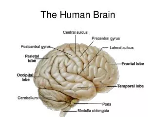
The human brain. Option E.5. Assessment statements. E.5.1 Label, on a diagram of the human brain, the medulla oblongata, cerebellum, hypothalamus, pituitary gland and cerebral hemispheres. E.5.2 Outline the function of each of the parts of the brain listed above.

E N D
The human brain Option E.5
Assessment statements • E.5.1 Label, on a diagram of the human brain, the medulla oblongata, cerebellum, hypothalamus, pituitary gland and cerebral hemispheres. • E.5.2 Outline the function of each of the parts of the brain listed above. • E.5.3 Explain how animal experiments, lesion and fMRI (functional magnetic resonance imaging) scanning can be used in the identification of the brain part involved in specific functions. • E.5.4 Explain sympathetic and parasympathetic control of the heart rate, movements of the iris and flow of the blood to the gut. • E.5.5 Explain the pupil reflex. • E.5.6 Discuss the concept of brain death and the use of the pupil reflex in testing for this. • E.5.7 Outline how pain is perceived and how endorphins can act as painkillers.
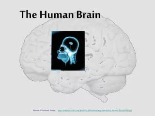
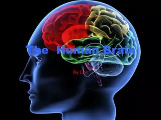
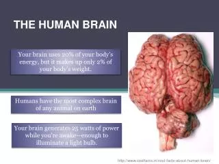
Hypothalamus maintains homeostasis, coordinating the nervous and the endocrine systems, secreting hormones of the posterior pituitary, and releasing factors regulating the anterior pituitary. Cerebral hemispheres act as the integrating center for high complex functions such as learning, memory and emotions. Cerebellum has two hemispheres and a highly folded surface. It coordinates unconscious functions, such as movement and balance. Pituitary gland has two lobes. The posterior lobe stores and releases hormones produced by the hypothalamus and the anterior lobe. It also produces and secretes hormones regulating many body functions. Medulla oblongata controls automatic and homeostatic activities, such as swallowing, digestion, vomiting, breathing, and heart activity.
Identification of brain parts involved in specific functions
Brain lesions area of tissue that has been damaged through injury or disease
Right and left hemispheres • Brain divided into right and left hemispheres • Connected by a thick band of axons called the corpus callosum • Left hemisphere • Contains areas important for communication • If damaged, person may have difficulty speaking or doing complicated movements • Right hemisphere • Specializes in receiving and analyzing information which comes in through all of our senses • If damaged, person may have difficulty identifying faces and locating an object correctly in space or even identifying melodies
Early experiments • Mid 1800s: Neurologists observed that people who had injuries on the left side had speech and language problems • People who had injuries in the same areas but on the right side of the brain had no language problems • Two areas of brain important for language are named for those neurologists: • Injury to Brocca’s interferes with the ability to volcalize words • Injury to Wernicke’s area affects the ability to put words into sentences
1960s: group of scientists interested in patients who had undergone surgery to sever their corpus callosum to relieve symptoms of epilepsy (the optic chiasma remains intact) • The Split Brain Experiment
fMRI uses radio waves and a strong magnetic field, not X-rays • Enables scientists to see the blood flow in the brain as it is occurring • Makes movies of what is going on in the brain as the subject performs tasks or is exposed to various stimuli • Can determine with some precision when regions of the brain become active and how long they remain active
fMRI used by doctors to determine: • A plan for surgery • Treatment for a stroke • Placement of radiation therapy for a brain tumor • Effects of degenerative brain disease such as Alzheimer’s • Diagnosing how a diseased or injured brain is working
Animal Experiments • Expose animals to addictive substances in controlled situations • Respond similarly to human: • Want more and more of the substance • Spend lots of time and energy getting it • Keep taking it despite adverse conditions • Have withdrawal symptoms on withdrawal of substance • Go back to the substance when stressed • Go back to the substance with another exposure to that substance
Animal model for addiction? • Animal is trained to press a lever to get a reward • Animal is given an injection of the addictive substance as it pushes the lever • Two levers available: one gives substance, one does not • If substance is reinforcing, animal will seek to repeat the experience by pushing that lever much more frequently and therefore, support the hypothesis that substance is addictive
Animal experiments can help us to determine way in which drugs promote abuse • Animal experiments cannot replicate the complete interaction of humans and drugs • Social factors can play a role • Addiction studies
Sympathetic and parasympathetic control • Peripheral nervous system considered in two parts, somatic system and autonomic system • Somatic system takes sensory information from sensory receptors to the CNS and then sends back motor commands from the CNS to the muscles • Autonomic system is involuntary and regulates activities of the glands, smooth muscle, and the heart. • Sympathetic system • Parasympathetic system
Antagonistic systems • Sympathetic system associated with fight or flight • You need quick energy • System increases heart rate, stroke volume to supply more glucose and oxygen • Dilates bronchi to give more oxygen • Dilates pupil by contracting radial muscles surrounding iris • Blood to gut is restricted by contraction smooth muscle of blood vessels carrying blood there
Parasympathetic takes over in a relaxed state • Nerves return the system to normal • Pupil of eye constricts • Heart rate slows, stroke volume is reduced • Blood returns to the digestive system • Smooth muscle of the blood vessels relax
Pupil reflex • Close your eyes and then suddenly open them • Pupil will close in response to the sudden input of light as the eyes open • Cranial reflex • Iris contains two sets of smooth muscle to open and close the pupil • Response caused by acetylcholine • Atropine stops the action of acetylcholine
Pathway of the pupil reflex • Optic nerve receives the messages from the retina in the back of the eye • Optic nerve connects with the pretectal nucleus of the brain stem • From the pretectal nucleus, a message is sent to the Edinger-Westphal nucleus whose axons run along the oculomotor nerves back to the eye • Oculomotor nerves synapses on the ciliary ganglion • Axons of the ganglion stimulate the circular muscle of the iris so it contracts
Brain death • Def: that time when a physician has determined that the brain and brain stem have irreversibly lost all neurological function • Patients in a coma have neurological signs that can be measured
Examinations for brain death includes checking: • Movement of extremities – if arms and legs are raised and let fall, there must be no other movement or hesitation in the fall • Eye movement – eyes must remain fixed showing lack of brain-to-motor-nerve reflex (as the head is turned there is no rolling motion of the eyes) • Corneal reflex – this must be absent (when a cotton swab is dragged over the cornea, the eye does not blink) • Pupil reflex – this must be absent (pupils do not constrict in response to a very bright light shone into both eyes) • Gag reflex – this must be absent (insertion of a small tube into the throat of a comatose patient will cause a gag reflex) • Respiration(breathing) response – this must be absent (if the patient is removed from a ventilator, the dead brain gives no response)
After being declared brain dead • Can still have spinal reflexes such as the knee jerk reflex • Spinal reflexes do not involve the brain • A short reflex motion can still be exhibited if the hand or foot is touched in a certain manner • Further tests: • Electroencephalogram (EEG) • Cerebral blood flow (CBF)
EEG • Measures brain activity in microvolts • Very sensitive test • Some electrical activity is shown on the EEG if a patient is in a deep coma • Life after death
CBF • Radioactive isotope is injected into the bloodstream • Radioactive counter is then placed over the head for about 30 minutes • If no activity is detected, this is conclusive evidence of brain death
Perception of pain • Pain signals are carried by peripheral nerve fibers from all over the body to the spinal cord and relayed to the sensory area of the brain • Peripheral fibers connect with pain receptors called nocioreceptors • Nocioreceptors are capable of sensing excess heat, pressure or chemicals from injured tissues • Nocioreceptors are located in the skin and also in the muscle, bones, joints and membranes around your organs • Nerve impulses of pain travel to the spinal cord • Ascending tracts in the spinal cord send the messages up to the brain
Response of pain by cerebral cortex • Can tell the muscles to stop the action which is causing the pain stimulus • Can alert the autonomic nervous system if the pain requires change in heart rate or breathing • Can direct other brain cells to release pain-suppressing endorphins
endorphins • First discovered by scientists studying opium addiction • Found receptors for the opiates, morphine and heroin in brain cells • Scientists found that the molecules made by plants were mimicking endorphins • Endorphins are CNS neurotransmitters with pain-relieving properties • Small peptides which bind to opiate receptors and block the transmission of impulses at synapses involved in pain perception