Download
1 / 63
720 likes | 1.29k Views
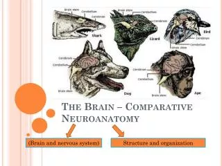
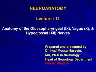
NEUROANATOMY Lecture : 3 Anatomy of the Brain Stem. Prepared and presented by: Dr. Iyad Mousa Hussein, Ph.D in Neurology Head of Neurology Department Nasser Hospital. LECTURE OBJECTIVES:. 1. Morphological Subdivisions of the Brain. 2. Site, Parts, and Functions of the Brain Stem.

E N D
NEUROANATOMY Lecture : 3 Anatomy of the Brain Stem Prepared and presented by: Dr. Iyad Mousa Hussein, Ph.D in Neurology Head of Neurology Department Nasser Hospital
LECTURE OBJECTIVES: 1. Morphological Subdivisions of the Brain. 2. Site, Parts, and Functions of the Brain Stem. 3. Parts of the Medulla Oblongata. 4. Connection, External, and internal Features of the Medulla Oblongata. 5. Connection, External, and internal Features of the Pons. 6. Connection, External, and internal Features of the Midbrain. 7. Location, Connections, and Functions of the Reticular Formation of the Brain Stem. 8. Motor and sensory pathway.
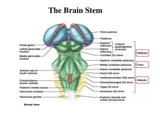
Morphological Subdivisions of the Brain • The Cerebrum:formed of right and left cerebral hemisphere. • 2. The Cerebellum:below the posterior part of the • cerebrum. • 3. The Brain Stem:formed of the following parts (from • downward): • a. Medulla oblongata. • b. Pons. • c. Midbrain.
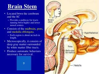
The Brain Stem • The brain stem lying infront of the cerebellum and • occupies the posterior cranial fossa. • The brain stemformed of the following parts (from • downward): • 1. Medulla oblongata. • 2. Pons. • 3. Midbrain.
Functions of the Brain Stem 1. It serves as a conduit for the ascending and descending tracts. 2. It contains important reflex centers associated with the control of cardiovascular, respiratory systems and consciousness. 3. It contains the important nuclei of cranial nerves III through XII.
The Medulla Oblongata The medulla oblongata is the lower part of the brain stem (3 cm). Extent: Above:it is continuous with pons. Below:it is continuous with the spinal cord at the foramen magnum.
Parts of the Medulla Oblongata Closed Medulla:it is the lower half of the medulla, as it encloses a central canal continuous with that of spinal cord. Open Medulla: it is the upper half of the medulla, as it opens into the fourth ventricle.
External Features of the Medulla Oblongata A. Anterolateral Surface: 1. The anterior median fissure. 2. The pyramid: formed by the pyramidal (corticospinal) tract. 3. The olive: formed by the inferior olivary nucleus. 4. The anterolateral sulci. 5. The posterolateral sulci: gives exit to the glossopharyngeal and vagus nerves. 6. The inferior cerebellar peduncle.
B. The posterior surface of the medulla oblongata: • 1. The posterior surface of the upper half (open • medulla):presents the following features from • medial to lateral: • a. The median longitudinal fissure. • b. Inferior fovea. • c. Hypoglossal trigone (triangle). • d. Vagal trigone. • d. Vestibular trigone. • 2. The posterior surface of the lower half (closed • medulla):presents the following features from • medial to lateral: • a. Posterior median fissure. • b. Gracile tract. • c. Cuneate tract.
Internal Structures of the Medulla Oblongata The main nuclei of the medulla oblongata: 1. Gracile nucleus:proprioceptive and fine touch from the lower 1/2 of the body. 2. Cuneate nucleus:proprioceptive and fine touch from the upper 1/2 of the body. 3. Inferior olivary nucleus:extrapyramidal function. 4. Inferior salivary nucleus:parasympathetic function via the glossopharyngeal nerve.
5. Spinal nucleus of trigeminal nerve:pain and temperature sensations from the face and scalp via the trigeminal nerve. 6. Solitary nucleus:taste sensations via the facial, glossopharyngeal and vagus nerves. 7. Nucleus ambiguous:pyramidal and extrapyramidal functions via motor fibers of the glossopharyngeal, vagus and accessory nerves. 8. Dorsal nucleus of vagus nerve:parasympathetic via the glossopharyngeal and vagus nerves. 9. Hypoglossal nucleus:motor function of the tongue via hypoglossal nerve.
Blood Supply of the Medulla Oblongata • The medulla oblongata is supplied by: • The vertebral arteries. • The anterior spinal artery. • The posterior spinal arteries. • The posterior inferior cerebellar arteries. • The basilar artery.
The Pons Extent: it extends from the medulla oblongata below to midbrain above and lies infront of the cerebellum.
External features of the pons A. The anterior surface of the pons: presents the following features: 1. The basilar groove: for basilar artery. 2. The transverse streaks: to form middle cerebellar peduncle. 3. The trigeminal nerve. 4. The middle cerebellar peduncle. 5. The abducent nerve. 6. The facial nerve. 7. The vestibule-cochlear nerve.
B. The posterior surface of the pons:presents the following features: 1. The median longitudinal sulcus: the middle line. 2. The medial eminence: produced by the abducent nucleus. 3. The facial colliculus: it produced by the facial nucleus. 4. The medullary stria: transverse nerve fibers which separate the posterior surface of pons from that of medulla oblongata.
Internal structures of the pons • Pontine nuclei: they form part of the cortico-ponto- • cerebellar pathway. • 2. Transverse fibers: pontocerebellar fibers. • 3. Longitudinal fibers: which include pyramidal and cortico- • pontine fibers. • 4. Nuclei of the trigeminal nerve: • a. Motor nucleus. • b. Sensory nucleus. • 5. Nucleus of the abducent nerve. • 6. Nuclei of the facial nerve: • Motor nucleus. • b. Superior salivary nucleus: parasympathetic function.
7. Nuclei of the vestibulocochlear nerve. 8. Lateral lemniscus: it is a band of ascending fibers carrying hearing impulses from both ears. 9. Spinal lemniscus: it is a band of ascending fibers carrying pain, temperature and crude touch from the opposite side of the body below the head. 10. Trigeminal lemniscus: it is a band of ascending fibers carrying pain, temperature and touch and proprioception from the opposite side of the face and scalp. 11. Medial lemniscus: it is a band of ascending fibers carrying proprioceptive and fine touch from the opposite side of the body below the head.
Blood Supply of the Pons • The pons is supplied by: • The anterior inferior cerebellar arteries. • The superior cerebellar arteries. • The basilar arteries.
The Midbrain Extent: It extends between the pons below and the diencephalon above. Connection: It connects pons and cerebellum to the subthalamic region of the diencephalon.
External features of the midbrain • A. The anterior surface of the midbrain:presents the following features: • 1. Two cerebral peduncles. • 2. Oculomotor Nerve: emerging from the medial • side of the cerebral peduncle.
B. The posterior surface of the midbrain:presents the following features: 1. Four colliculi (rounded elevations): which include: a. Two superior colliculi (Visual function). b. Two inferior colliculi (Auditory function). 2. Two superior cerebellar peduncles. 3. Superior medullary velum (membrane): lies between the two superior cerebellar peduncles. 4. The trochlear nerve. C. The lateral surface of the midbrain: presents the following features: 1. Brachium of superior colliculus. 2. Brachium of inferior colliculus.
Internal Structures of the Midbrain In transverse section of the midbrain, the Sylvius aqueduct divides it into two main parts: 1. Tectum:the smaller dorsal part behind the Sylvius aqueduct, and consists of: a. Two Superior Colliculi: reflex centers of vision. b. Two Inferior Colliculi: reflex centers of hearing. 2. Two cerebral peduncles: the larger ventral part infront of aqueduct, it consists of three parts: a. Crus cerebri: the most anterior part which consists of pyramidal and cortico-nuclear fibers. b. Substantia Nigra: a thick lamina of gray mater. It is an extrapyramidal center. c. Tegmentum: the posterior part of the cerebral peduncle. It contains ascending tracts, certain nuclei, reticular formation of the midbrain and decussation of the superior cerebellar peduncle.
Internal Structures of the Midbrain The main nuclei of the midbrain: 1. Nucleus of oculomotor nerve. 2. Nucleus of the trochlear nerve. 3. Red nucleus: it is an important extrapyramidal centre. 4. Mesencephalon nucleus of the trigeminal nerve: it receives proprioceptive impulses from the masticator and ocular muscles. 5. Inferior colliculus: it is a centre for auditory reflexes. 6. Superior colliculus: it is a centre for visual reflexes. 7. Substantia nigra: it is an extrapyramidal centre.
Transverse section of the midbrain at the level of the superior colliculus
Transverse section of the midbrain at the level of the inferior colliculus