Download

1 / 48
651 likes | 1.64k Views
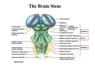
The brain stem. SHANDONG UNIVERSITY Liu Zhiyu. The brain stem. Consists of Midbrain Pons Medulla oblongata. The brain stem. Position The brain stem occupies the posterior cranial fossa of the skull. Gross Appearance of the Brain Stem.

E N D
The brain stem SHANDONG UNIVERSITY Liu Zhiyu
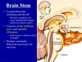
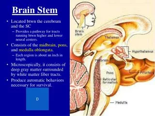
The brain stem Consists of • Midbrain • Pons • Medulla oblongata
The brain stem Position • The brain stem occupies the posterior cranial fossa of the skull
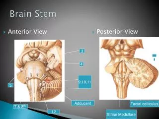
Ventral Surface of the Brain Stem Medulla oblongata • Pyramid: Composed of bundles of nerve fiber, pyramidal tract • Decussation of pyramid: Formed by crossing fibers of pyramidal tract • Olive: Produced by underlying inferior olivary nucleus • Anterolateral sulcus: Emerge the roots of hypoglossal nerve • Retroolivary sulcus: Emerge the roots of glossopharyngeal, vagus and accessory nerves
Ventral Surface of the Brain Stem Pons • Basilar part • Basilar sulcus • Bulbopontine sulcus: There emerge, from medial to lateral, the abducent, facial and vestibulocochlear nerves • Middle cerebellar peduncle • Trigeminal nerve Emerges from the junction of basilar part and middle cerebellar peduncles • Pontocerebellar trigone: The junction of medulla, pons and cerebellum
Ventral Surface of the Brain Stem Midbrain • Crus cerebri • Interpeduncular fossa Oculomotor nerve emerges on the medial side crus cerebri • Posterior perforated substance
Dorsal Surface of the Brain Stem Medulla oblongata • Lower portion • Gracile tubercle:produced by underlying gracile nucleus • Cuneate tubercle:produced by underlying cuneate nucleus • Inferior cerebellar peduncle • Obex • Upper portion:forms the lower half of rhomboid fossa
Dorsal Surface of the Brain Stem Pons • Superior cerebellar peduncle • Superior medullary velum • Trochlear nerve
Dorsal Surface of the Brain Stem Midbrain • Superior colliculus Constitute centers for visual reflexes • Inferior colliculus Associated with auditory pathway • Brachium of superiorcolliculus • Brachium of inferior colliculus
Fourth ventricle Central canal →fourth ventricle →mesencephalic aqueduct→third ventricle Position • Situated ventral to cerebellum, and dorsal to pons and cranial half of medulla
Floor of the Fourth ventricle — Rhomboid Fossa Boundaries • Inferolateral: gracile and cuneate tubercles, inferior cerebellar peduncle • Superolateral: superior cerebellar peduncle • Lateral recess
Floor of the Fourth ventricle — Rhomboid Fossa Features • Median sulcus • Sulcus limitans • Vestibular area Overlies vestibular nuclei • Acoustic tubercle Overlying dorsal cochlear nucleus • Medial eminence • Striae medullares
Floor of the Fourth ventricle — Rhomboid Fossa • Facial colliculus Overlies nucleus of abducent n. and genu of facial nerve • Hypoglossal triangle Overlies hypoglossal nucleus • Vagal triangle Overlies dorsal nucleus of vagus nerve • Funiculus separans • Area postrema • Locus ceruleus Bluish-gray in color, it owes its color to a group of deeply pigmented nerve cells
Roof of the Fourth ventricle • Anterior part: Formed by superior cerebellar peduncle and superior medullary velum • Posterior part: Formed by inferior medullary velum and choroid plexus of fourth ventricle • Three apertures • Median aperture of fourth ventricle • Two lateral apertures of fourth ventricle
Internal structure of the brain Stem General somatic motor nuclei Special visceral motor nuclei • Central canal • Gray matter • Cranial nerve nuclei • Relay nuclei • White matter • Reticular formation of brain stem General visceral motor nuclei Visceral sensory nuclei( general and special ) General somatic sensory nuclei Special visceral motor nuclei
General somatic motor nuclei • Nucleus of oculomotor n. • Nucleus of trochlear n. • Nucleus of abducent n. • Nucleus of hypoglossal n.
General somatic motor nuclei • Nucleus of oculomotor n. • Nucleus of trochlear n. • Nucleus of abducent n. Nucleus of hypoglossal n.
Special visceral motor nuclei • Motor nucleus of trigeminal n. • Nucleus of facial n. • Nucleus ambiguus • Accessory nucleus
Special visceral motor nuclei • Motor nucleus of trigeminal n. • Nucleus of facial n. • Nucleus ambiguus • Accessory nucleus
General visceral motor nuclei • Accessory oculomotor nucleus • Superior salivatory nucleus • Inferior salivertory nucleus • Dorsal nucleus of vagus n.
Visceral sensory nuclei ( general and special ) • Nucleus of solitary tract
Visceral sensory nuclei ( general and special )
General somatic sensory nuclei • Mesencephalic nucleus of trigeminal n. • Pontine nucleus of trigeminal n. • Spinal nucleus of trigeminal n.
Special somatic sensory nuclei • Cochlear nuclei • Vestibular nuclei
Relay nuclei • Superior colliculus • Nucleus of inferior colliculus • Red nucleus • Substantia nigra • Pretectal area • Superior olivery nucleus • Pontine nucleus • Nucleus ceruleus • Gracile nucleus • Cuneate nucleus • Inferior olivary nucleus
White matter Ascending tracts • Medial lemniscus • Spinothalamic tract(Spinal lemniscus ) • Trigeminothalamic tract(Trigeminal lemniscus ) • Lateral lemniscus
Ascending tracts Central thalamic radiation Medial lemniscus VPL 3°neurons Medial lemniscus Gracile and cuneate nuclei 2°neuron Decussation of medial lemniscus Fasciculus cuneatus T4 Fasciculus gracilis Spinal ganglion 1°neuron
Ascending tracts Central thalamic radiation Spinothalamic tract (Spinal lemniscus) VPL 3°neurons Spinal lemniscus anterior spinothalamic tract LaminaⅠ,Ⅳ~Ⅶ 2°neuron Lateral spinothalamic tract Spinal ganglia 1°neuron
Ascending tracts Central thalamic radiation Trigeminothalamic tract (Trigeminal lemniscus) VPM 3°neurons Pontine nucleus of V 2°neuron Trigeminal lemniscus Trigeminal ganglion 1°neuron Spinal tract of trigeminal n. Spinal nucleus of V 2°neuron
Ascending tracts Lateral lemniscus
Descending tracts • Pyromidal tract • Corticospinal tract • Corticonuclear tract • Rubrospinal tract • Tectospinal tract • Vestibulospinal tract • Reticulospinal tract
Reticular Formation of Brain Stem • Ascending reticular activating system (ARAS) • Motor central and vital centres • Reticulospinal tract • Cardiovascular center • Respiratory center • Serotonergic rapheal nuclei
Medulla oblongata Lower part (closed part) • Two decussations • Decussations of medial lemniscus • Decussations of pyramid
Medulla oblongata Upper part (open part) • Appearance of inferior olivary nuculeus and inferior cerebellar peduncle • Enlargement of central canal to form the fourth ventricle floor
Pons • Tegmentum of pons Directed upward continuation of medulla oblongata • Basilar part Contain both longitudinal and transverse fibers intermixed with pontine nuclei
Midbrain Rectum of midbrain Includes superior and inferior colliculi Cerebral peduncle • Tegmentum contain ascending tracts, mesencephalic aqueduct, central gray matter, recticular formation and so on • Substentia nigra • Crus cerebri • Pyramidal tractmiddle three-fifths of the crus • Frontopontine tractmedial one-fifth • Pariatotempopontine tractlateral one-fifth
Midbrain Rectum of midbrain Includes superior and inferior colliculi Cerebral peduncle • Tegmentum contain ascending tracts, mesencephalic aqueduct, central gray matter, red nucleus, recticular formation and so on • Substentia nigra • Crus cerebri • Pyramidal tractmiddle three-fifths of the crus • Frontopontine tractmedial one-fifth • Pariatotempopontine tract • lateral one-fifth