Download
1 / 83
830 likes | 834 Views
Learn about the Central Nervous System, Peripheral Nervous System, and Neurons. Understand the functions and responses of the Autonomic Nervous System. Discover the fascinating concept of Mirror Neurons.

E N D
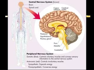
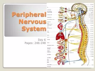
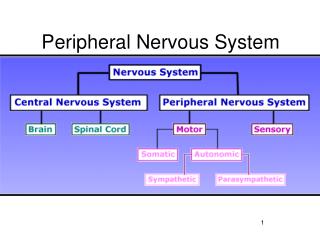
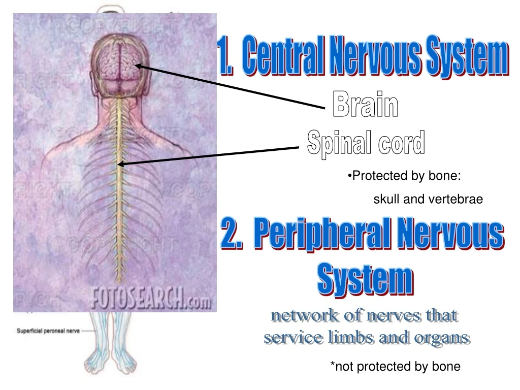
1. Central Nervous System Brain Spinal cord • Protected by bone: skull and vertebrae 2. Peripheral Nervous System network of nerves that service limbs and organs *not protected by bone
Peripheral Nervous System A. Somatic NS Controls VOLUNTARY body movements, through the action of skeletal muscles. Interacts with surroundings using senses B. Autonomic NS Maintains INVOLUNTARY actions such as heart rate, digestion, sweating, sexual arousal 1. sympathetic: Vigorous activity. Ex: “Fight or Flight” response parasympathetic: 2. Non-emergency activity. Ex: “Rest and Digest”
Examples: Rate decreased Force decreased Broncial muscle contracted Pupil contstricted Digestion increased Sphincter relaxed Increase urine secrestion
Parasympathetic Responses • Enhance “rest-and-digest” activities • Mechanisms that help conserve and restore body energy during times of rest • Normally dominate over sympathetic impulses • SLUDD type responses = salivation, lacrimation, urination, digestion & defecation and 3 “decreases”--- decreased HR, diameter of airways and diameter of pupil • Paradoxical fear when there is no escape route or no way to win – causes massive activation of parasympathetic division – loss of control over urination and defecation Sympathetic Responses Dominance by the sympathetic system is caused by physical or emotional stress -- “E situations” – emergency • embarrassment • excitement • exercise • Alarm reaction = flight or fight response – dilation of pupils – increase of heart rate, force of contraction & BP – decrease in blood flow to nonessential organs – increase in blood flow to skeletal & cardiac muscle – airways dilate & respiratory rate increases – blood glucose level increase
Sympathetic response: Lexus Commercial https://www.youtube.com/watch?v=qhMeHPwyJNI
The Neuron (nerve cell): Functional unit of the Nervous System
Neuron: nerve cell dendrites Cell body (soma) axon Myelin sheath (orange padding of the Schwann Cell) Synapse axon Node of Ranvier (gap)
Dendrites Soma Nucleus Nucleolus Axon Node of Ranvier Schwann Cell Axon terminal (ending)
Motor Neuron Sensory Neuron Dendrite Axon Soma “Afferent” : Ascending “Efferent”: Descending Sensory neuron receives information from the senses (environment) and sends it to the CNS A motor neuron receives information from the CNS and sends it to a muscle or gland.
Axon terminal Mylein Sheath Cell Body Dendrites Axon Node of Ranvier)
Sensory impulse Sensory neurons Receptor Motor neurons Optic: eye Auditory: ear Olfactory: smell Taste buds: taste Touch: skin receptors Motor impulse Effector Muscle gland
2 Additional neurons 1. Interneuron • Found in the CNS only. • They associate or “connect” sensory neurons and motor neurons. 2.Mirror Neurons • allow humans to “mimic” each other • possibly allow us to feel empathy • New field of study
The Neurons that Shaped Civilization Lecture on Mirror Neurons (7min) http://www.ted.com/talks/vs_ramachandran_the_neurons_that_shaped_civilization.html http://ftw.usatoday.com/2017/10/yankees-gary-sanchez-baseball-to-the-groin-oof-wild-card-mlb PBS; Mirror Neurons (15 min)
Mirror neurons are found all over the brain and they look just like other neurons. What makes them special is the web of connections that link these neurons in the motor and sensory systems to the limbic centers that process visceral and emotional reactions.
While they may be in place at birth, they are vastly expanded through experience. A baby smiles…her mother smiles back…the brain sets up a circuit.
Evidence for mirror neurons: When a researcher would pick up raisins or sunflower seeds, neurons that a macaque would use when engaged in the same task would fire. In humans, the same neurons fired when subjects felt a glove brush their leg and when they watched a video of an actor’s leg being brushed by a glove. The thought of a loved one’s hand receiving an electric shock lights up many of the same brain areas as shocks that are directly experienced.
Can even indicate strength of emotion… The same mirror neurons fired when the subjects saw a hand reaching for both of these, BUT… Neurons fired strongly neurons fired more weakly
Questions • The brain and spinal cord make up the _______________________ • Which of the following do not belong in this grouping • Autonomic – sympathetic – parasympathetic – central nervous system • Stimuli from the environment are received by _______________ • The insulating sheath of the axon is composed of ________________ • Impulses that go to the brain are? Ascending or Descending • The gaps between the myelin in an neuron are called? ________________ • The gaps that exist between two neurons are called _____________________ • The part of the neuron where the nucleus is found is the __________________ • What is “fight or flight”? • What part of the nervous would be responsible for flight or flight? • Explain what else happens to your body during this response.
Synapse • Point of communication between two nerve cells. • Three parts: • presynaptic membrane • synaptic cleft (gap) • postsynaptic membrane neurotransmitter • A __________________is the chemical that is exchanged at the synapse. This allows the chemical message to be______________________________ Passed from one neuron to the next
Action Potential http://highered.mcgraw-hill.com/sites/0072495855/student_view0/chapter14/animation__the_nerve_impulse.html https://www.youtube.com/watch?v=Ibzfwtdtong&feature=related#t=196.3041374 Synapse http://highered.mcgraw-hill.com/sites/0072495855/student_view0/chapter14/animation__chemical_synapse__quiz_2_.html http://www.youtube.com/watch?v=90cj4NX87Yk&feature=related
Brain Plasticity (video clip) Nerve Proliferation and Pruning: “use it or lose it” in the brain. Babies have about 15,000 connections per neuron while adults have about a third less (10,000). Children between age 1-2 can make up to 2 million neurological connections per minute. The number of cells does not change, only the number of connections! Although plasticity continues throughout life, the brain undergoes two major developmental phases, one in the womb and the second during the childhood/teen years. Links that are used are reinforced and strengthened while the ones that aren’t used are “pruned” or die off.
Brain Plasticity: Your Brain Changes • Chemical: neurotransmitters act on the synapse and drive behavior These chemical signals create neural pathways and are correspond to immediate behavior and have short term consequences (short term memory). analogy: acetylcholine causes a muscle to contract • Structural: Continued and sustained chemical firing causes structural • changes to the brain. • analogy: sustained muscle contractions over time causes the muscle to change in structure (i.e. it gets bigger) 3. Functional: Continued and sustained chemical firing causes structural changes to the brain. ex: sustained muscle contractions over time causes the muscle to change in structure (ie: gets bigger) • Plasticity varies from person to person (maybe genetic?) • Plasticity is behavior driven…dictated by what you do and what you do not do. • Drug addiction
Central Nervous System Peripheral Nervous System Somatic Autonomic Sympathetic Parasympathetic
Spinal Nerves: 31 pairs of nerves leave the spinal cord and carry impulses to and from the rest of the body (except for the head which is served by the cranial nerves). • 8 pairs of cervical • 12 pairs of thoracic • 5 pairs of lumbar • 5 pairs of sacral • 1 pair of coccygeal
Forebrain * Largest part of the brain • Cerebrum (cerebal cortex) • Thalamus • Pituitary Also: • Limbic System • Hypothalamus (regulate homeostasis) • Hippocampus (build new memories) • Amygdala (emotional responses) • fear, anger, aggression, sexual Limbic System
Main Parts of the Limbic System • Hypothalamus • Hippocampus • Amygdala
Hindbrain Consists of: • Cerebellum • Pons • Medulla oblongata
Midbrain • Function: • Relays visual and auditory inputs as they enter the brain
The External Brain Parietal Lobe Frontal Lobe Occipital Lobe Temporal Lobe Cerebellum Brainstem * There are two hemispheres in the brain: Left and Right. Which one is shown in this diagram? __________________ RIGHT
Left Hemisphere controls RIGHT side of the body Right Hemisphere controls LEFT side of the body
Frontal Lobe: Functions: • How we know what we are doing within our environment (Consciousness). • How we initiate activity in response to our environment = reasoning / thinking • Controls our impulsive responses. • Controls our expressive language. • Controls motor functions. • Involves word associations. • Memory for habits and motor activities. Observed Problems: • Loss of simple movement of various body parts (Paralysis). • Inability to plan a sequence of complex movements. • Loss of spontaneity in interacting with others. • Loss of flexibility in thinking. • Persistence of a single thought (Perseveration). • Inability to focus on task (Attending). • Mood changes (Emotionally Labile). • Changes in social behavior. • Changes in personality. • Difficulty with problem solving. • Inability to express language (Broca's Aphasia). Frontal Lobe
Parietal Lobe Function • Location for visual attention. • Location for touch perception. • Goal directed voluntary movements. Manipulation of objects. • Integration of different senses that allows for understanding a single concept. Observed Problems: • Inability to attend to more than one object at a time. • Inability to name an object. • Inability to locate the words for writing. • Problems with reading (Alexia). • Difficulty with drawing objects. Difficulty in distinguishing left from right. • Difficulty with doing mathematics (Dyscalculia). • Lack of awareness of certain body parts and/or surrounding space (Apraxia) that leads to difficulties in self-care. • Inability to focus visual attention. . Parietal Lobe
Occipital Lobe: Location: Most posterior lobe, at the back of the head. Functions: • Vision • Color recognition Observed Problems: • Defects in vision (Visual Field Cuts). • Difficulty with locating objects in environment. • Difficulty with identifying colors (Color Agnosia). • Production of hallucinations • Visual illusions - inaccurately seeing objects. • Word blindness - inability to recognize words. • Difficulty in recognizing drawn objects. • Inability to recognize the movement of an object (Movement Agnosia). • Difficulties with reading and writing Occipital Lobe
Temporal Lobe: Location: Side of head above ears. Functions: • Hearing ability • Memory acquisition • Some visual perceptions • Categorization of objects. Observed Problems: • Difficulty in recognizing faces • Difficulty in understanding spoken words (Wernicke's Aphasia). • Disturbance with selective attention to what we see and hear. • Difficulty with identification of, and verbalization about objects. • Short-term memory loss. • Interference with long-term memory • Increased or decreased interest in sexual behavior. • Inability to categorize objects. • Right lobe damage can cause persistent talking. • Increased aggressive behavior Temporal lobe
Summary Frontal Parietal Occipital Temporal Cognition Information processing Pain/touch sensation Spatial orientation Speech Visual perception Motor functions Problem solving Planning Reasoning Judgment Impulse memory Vision Color recognition Emotional response Hearing Memory Speech
Gyrus and sulcus The convolutions of the brain are made up of “ridges and grooves”
Speech and Language Broca’s area Wernicke’s Area “Language output”. Responsible for spoken language. People with damage to this area have trouble creating works and sentences. • Motor speech. “Language input”. Responsible for language comprehension. People with damage to this area can speak, but it is incoherent. • Sensory speech * These parts are found in the left hemisphere only!!
Broca's Area Motor Speech Wernicke's Area Sensory speech
Cerebellum: Location: at the base of the skull Functions: • Coordination of voluntary movement • Balance and equilibrium • Some memory for reflex motor acts. Observed Problems: • Loss of ability to coordinate fine movements. • Loss of ability to walk. • Inability to reach out and grab objects. • Tremors. • Dizziness (Vertigo). • Slurred Speech (Scanning Speech). • Inability to make rapid movements cerebellum