Download
1 / 38
380 likes | 512 Views
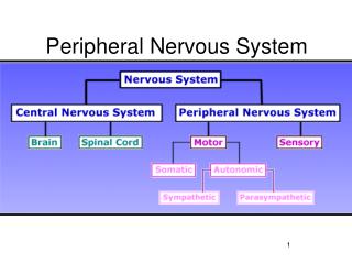
Peripheral Nervous System. Chapter 14. PNS. Made of the following: 12 CRANIAL NERVES 31 PAIRS OF SPINAL NERVES ALL SMALL BRANCHES FROM THE ABOVE GROUPS INCLUDES AFFERENT AND EFFERENT INFORMATION (information to and away from spinal cord. Spinal Nerves. 31 pairs Connected to spinal cord

E N D
Peripheral Nervous System Chapter 14
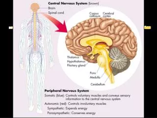
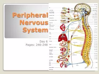
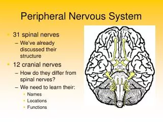
PNS • Made of the following: • 12 CRANIAL NERVES • 31 PAIRS OF SPINAL NERVES • ALL SMALL BRANCHES FROM THE ABOVE GROUPS • INCLUDES AFFERENT AND EFFERENT INFORMATION (information to and away from spinal cord
Spinal Nerves • 31 pairs • Connected to spinal cord • Cervical -8 pairs • Thoracic- 12 pairs • Lumbar- 5 pair • Sacral – 5 pair • Coccygeal- 1 pair • Nerves exit between vertebrae through the intervertebral foramen (IVF) • Each nerve has a ventral and dorsal root that joins before leaving the IVF
Nerve Plexus • After nerves leave the spinal cord they form complex networks of nerves called PLEXUSES. • CERVICAL PLEXUS (C1 nerve- C 4) • BRACHIAL PLEXUS (C 5- T1) • LUMBAR PLEXUS (L1-S4) • SACRAL PLEXUS (S5-Coccygeal 1) • **No particular plexus from the Thoracic region T2-T12
PLEXUS cont… • Cervical Plexus • Sensory to head and front of neck, upper shoulder, motor to neck muscles • Brachial Plexus • Lower Shoulder and entire arm • Lumbar Plexus • Supplies thigh and leg • Sacral and Coccygeal Plexus • Forms the sciatic nerve (largest nerve in the body)
Each nerve emerges from the spinal cord at points called ROOTS Dorsal Root Ganglion Ventral root ganglion
Divisions of the PNS • Somatic Motor System • Somatic reflexes • Reflex- the action that results from a nerve impulse passing over reflex arc. A predictable response to a stimulus. • Somatic reflexes involve glandular secretions or muscle contraction. • Autonomic reflexes involve contraction of smooth or cardiac muscle or secretion of glands.
Reflexes • Knee Jerk reflex (L2,3,4) • Ankle jerk reflex(S1,2) • Babinski reflex (plantar flexion when foot stroked)- cranial reflex • Corneal reflex (blinking )- cranial reflex • Abdominal reflex (T8-12)
Autonomic Nervous System • Efferent information from brain and SC to the visceral organs. • Depends on feedback mechanisms from sensory pathways. • Temperature regulation • Smooth muscles and glands
Sympathetic and parasympathetic systems (fig 14-17) • Sympathetic • Fight or flight responses- blood vessels dilate, heart rate increases, blood to muscles, pupils dilate, respiration increases- adrenaline rush • Sympathetic chain ganglion run the length of the spinal cord • Parasympathetic • Rest and digest- heart rate decreases. Visceral organs are active, body repairs itself. • Axons located in cranial nerves 3, 7, 9, 10 and some pelvic nerves.
9.15 Autonomic Nervous System Sympathetic - energy, high stress, emergency Fight or Flight Parasympathetic - resting, normal Divisions act antagonistically - one is exhitatory, other inhibits
Autonomic Neurotransmitters • Norepinephrine and acetylcholine. • Norepinephrine has alpha and beta receptors • Beta blockers are meds given to control irregular heart beats, high blood pressure etc.. These are cases where sympathetic system is in overdrive. • Acetylcholine (table 14-7 p 438) • Nicotinic receptors • Muscarinic receptors
Sense Organs Chapter 15 Sensory receptors are called sense organs. Classified by location 1.Externoreceptors- near the body surface temperature, pain, pressure and touch 2.Visceroreceptors –in body organs Info about pressure, stretch (stomach ), hunger, thirst etc… 3. Proprioception- In joint capsules and muscles give information about where the organ is in relation to the body
Classification by Stimulus • Mechanoreceptors- position • Chemoreceptors- chemical changes (ie.pH) • Thermoreceptors- temperature changes • Nociceptors- pain resulting from intense stimuli. (ie= toxic chemicals, loud noise, pressure etc…) • Photoreceptors- Only in the eye- light receptors
Classification by structure • Free Nerve endings • Simplest form of receptor. Abundant • Endings are dendritic knobs. • Pain, nociception- not found in the brain. Headaches are referred pain from other areas • Encapsulated Nerve endings • Touch and Pain receptors • Stretch receptors (muscle spindles) • Table 15-1 p452
Smell- Olfactory Receptors • Chemoreceptors in the basal membrane of the epithelium of the nasal passageway • Fibers of the olfactory bulb pass through the cribiform plate into the olfactory recess. • Smell can create long lasting memories. Dental office, baby smell, kitchen smell brings back memories
Taste- Gustatory stimuli Taste: Taste buds are very similar to smell. Sensory receptors in your taste buds act similar to the olfactory nerves and send the chemical signal to the brain. There are basically 5 receptor types. • Salty • Sour • Sweet • Bitter • Umami • The umami taste is that of monosodium glutamate and has recently been recognized as a unique taste, as it cannot be elicited by any combination of the other four taste types. Glutamate is present in a variety of protein-rich foods, and particularly abundant in aged cheese.
Olfactory (smell) bulb Taste sensory area Olfactory nerve Thalamus Cerebral cortex Smell receptor Nasal cavity Smell sensory area Taste bud Taste pore Taste receptor Sensory nerve fibers The Senses of Smell and Taste Section 35-4
Taste • Taste buds are the sense organs that respond to gustatory or taste stimuli. • Chemoreceptors- stimulated by chemicals and dissolved in saliva • Gustatory cells- specialized taste buds, gustatory hairs extend from the gustatory cell into taste pore. • Chemicals bind to G proteins or ion channels and messages are sent to the brain for interpretation, exact mechanism is unknown.
Sense of Hearing and Balance • 3 Parts to the Ear • External Ear • Auricle or Pinna – external ear • External auditory meatus- canal • Middle Ear (tympanic cavity) • 3 auditory ossicles • Tympanic membrane to round window • Inner Ear • Labryinth- bony and membranous • Bony labryinth- membranous semicircular canals • Contains endolymph and perilymph
Middle Ear • Hollowed out opening in the temporal bone • 3 ossicles • Malleus, incus and stapes • Opening into the Eustachian or auditory tube which connects to the throat. Leads to infections, especially in small children, when fluid drains into the cavity.
Inner Ear- aka Labyrinth • 3 parts: • Cochlea- hearing • vestibule, and semicircular canals- balance.
Vestibular Canals: Balance • Pp461-463 • Senses position of head relative to gravity or acceleration or deceleration of the body. Fluid filled canals.
Vision • Sclera- outer coat • Tough, white connective tissue • Cornea- the transparent anterior portion that lies over the iris. No blood vessels in the cornea • Canal of Schlemm- ring shaped venous sinus found deep within the anterior portion of the sclera it junction with the cornea. • Choroid- middle coat • Blood vessels and pigment • Includes IRIS
Vision • Retina- innermost coat of eye • Photoreceptor neurons- visual receptors • Rods- highly light sensitive • Cones- less sensitive need brighter lights. • Bipolar neurons • Ganglionic neurons • Cavities and humors • Cavities • Anterior- in front of lens • Posterior- behind iris, but in front of lens
Vision cont… • Humors • Aqueous humor- fills both chambers of the anterior cavity, clear watery • Vitreous humor- fills the posterior cavity. Semisolid material, helps maintain sufficient pressure. • Muscles • Extrinsic- change position of the eye • Intrinsic- control iris (Pupil), give shape to lens
Accessory structures to eye • Eyebrows and lashes- some protection • Eyelids • Lacrimal glands- secrete tears • Lacrimal canals • Lacrimal sacs • Nasolacrimal ducts
Sight • Forming image on Retina • Refraction of light rays • http://phet.colorado.edu/en/simulation/geometric-optics • Accommodation of lens- increasing curvature to achieve the greater refraction needed for near vision. • Constriction of pupil- control amount of light hitting retina- accommodates lens • Convergence of eye- movements of the 2 eyeballs inward so that their visual axes come together at the object viewed; the closer the object the greater the degree of convergence necessary to maintain single vision.
Rods and Cones • Rods- photopigment in rods is rhodopsin; highly light sensitive; breaks down into opsin and retinal; separation of opsin and retinal in the presence of light causes an action potential in rod cells; energy is needed to reform rhodopsin. Night vision • Cones- 3 types, with each having a different photopigment; cone pigments are less light sensitive than rhodopsin and need brighter light to breakdown.- color, bright light
Neuronal pathway of vision • Fibers that conduct impulses from the rods and cones reach the visual cortex in the occipital lobes via the optic nerves, optic chiasma, optic tracts and the optic radiations. • Optic nerve contains fibers from only one retina, but optic chiasma contains fibers from the nasal portion of both retinas; these anatomical facts explain peculiar visual abnormalities that sometimes occur