Download
1 / 19
190 likes | 295 Views
Effects of detraining on cardiovascular responses to exercise: role of blood volume EDWARD F. COYLE, MAR1 K. HEMMERT, AND ANDREW R. COGGAN J. Appl. Physiol. 60(l): 95-99, 1986.

E N D
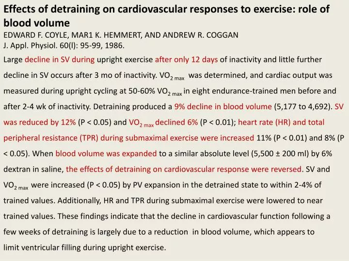
Effects of detraining on cardiovascular responses to exercise: role of blood volume EDWARD F. COYLE, MAR1 K. HEMMERT, AND ANDREW R. COGGAN J. Appl. Physiol. 60(l): 95-99, 1986. Large decline in SV during upright exercise after only 12 days of inactivity and little further decline in SV occurs after 3 mo of inactivity. VO2 max was determined, and cardiac output was measured during upright cycling at 50-60% VO2 max in eight endurance-trained men before and after 2-4 wk of inactivity. Detraining produced a 9% decline in blood volume (5,177 to 4,692). SV was reduced by 12% (P < 0.05) and VO2 max declined 6% (P < 0.01); heart rate (HR) and total peripheral resistance (TPR) during submaximal exercise were increased 11%(P < 0.01) and 8% (P < 0.05). When blood volume was expanded to a similar absolute level (5,500 ± 200 ml) by 6% dextran in saline, the effects of detraining on cardiovascular response were reversed. SV and VO2 max were increased (P < 0.05) by PV expansion in the detrained state to within 2-4% of trained values. Additionally, HR and TPR during submaximal exercise were lowered to near trained values. These findings indicate that the decline in cardiovascular function following a few weeks of detraining is largely due to a reduction in blood volume, which appears to limit ventricular filling during upright exercise.
Effect of Aerobic Exercise on Blood Pressure: A Meta-Analysis of Randomized, Controlled Trials Seamus P. Whelton; Ashley Chin, MPH, MA; Xue Xin, MD, MS; and Jiang He, MD, PhD Ann Intern Med. 2002;136:493-503.
This meta-analysis is a comprehensive examination of the effect of aerobic exercise on blood pressure and is based on randomized, controlled clinical trials. Our meta-analysis included 54 clinical trials that involved 2419 participants and were conducted in a wide range of geographic regions and ethnic populations. Our study showed that aerobic exercise has an impressive blood pressure–lowering effect: 3.84 mm Hg for systolic blood pressure and 2.58 mm Hg for diastolic blood pressure. It is important to distinguish between the individual and public health implications of our findings. The blood pressure reduction that we observed may be of moderate interest to practitioners treating individual patients. However, a small decrease in the population’s average blood pressure level should dramatically reduce incidence of and death from cardiovascular disease in communities. All forms of exercise seem to be effective in reducing blood pressure. Aerobic exercise reduces insulin resistance and insulin levels in hypertensive patients. Change in blood pressure during exercise is strongly associated with reduction in serum concentrations of total cholesterol and insulin resistance.
Effects of Aging, Sex, and Physical Training on Cardiovascular Responses to Exercise Takeshi Ogawa, MD; Robert J. Spina, PhD; Wade H. Martin III, MD; Wendy M. Kohrt, PhD; Kenneth B. Schechtman, PhD; John O. Holloszy, MD; and Ali A. Ehsani, MD (Circulation 1992;86:494-503) To investigate the mechanism of the age-related decline in exercise capacity, we measured oxygen uptake, cardiac output, heart rate, and other cardiovascular responses to submaximal and maximal treadmill exercise in healthy sedentary and endurance exercise-trained younger and older men and women. We estimated fat-free mass in the same individuals from measurements of body weight and density. Our findings provide evidence that the decline in VO2max with age is related primarily to a lower maximal cardiac output. Although a slower maximal heart rate accounts for a portion of this effect, a smaller stroke volume is of greater importance. Differences in VO2max between 25- and 65-year-old sedentary subjects of the same sex are approximately 40%. (10% per decade). After normalization of results to fat-free mass, however, VO2max , maximal cardiac output, and stroke volume were an average of 24%, 17%, and 8% lower, respectively, in older than in younger individuals.
The smaller stroke volume observed in older subjects at maximal exercise was associated with a higher mean blood pressure in women and sedentary men. For these groups, stroke work in the older subjects was equal to or greater than that in younger individuals. Stroke volume and cardiac output at maximal exercise were lower in women than in men, even after normalization to weight. Normalization of results to fat-free mass eliminated the sex difference entirely in sedentary subjects and substantially reduced it in trained individuals. Thus, the sex difference is largely a result of the greater percentage of body fat in women. However, there were sex differences in mechanisms by which exercise capacity was enhanced in conditioned versus sedentary subjects. Training status had a larger effect on stroke volume and maximal cardiac output but a smaller effect on maximal arteriovenous oxygen difference in men than in women. Sex differences in the nature and magnitude of adaptations to training were particularly evident in older subjects.
Exercise as Cardiovascular Therapy Roy J. Shephard, MD, PhD, DPE; Gary J. Balady, MD Circulation 1999;99;963-972 Possible Biological Mechanisms for Exercise-Induced Reductions in All-Cause and Cardiac Mortality Cardiovascular influences Reduction of resting and exercise heart rate Reduction of resting and exercise blood pressure Reduction of myocardial oxygen demand at submaximal levels of physical activity Expansion of plasma volume Increase in myocardial contractility Increase in peripheral venous tone Favorable changes in fibrinolytic system Increased endothelium-dependent vasodilatation Increased gene expression for nitric oxide synthase Enhanced parasympathetic tone Possible increases in coronary blood flow, coronary collateral vessels, and myocardial capillary density
Metabolic influences Reduction of obesity Enhanced glucose tolerance Improved lipid profile Lifestyle influences Decreased likelihood of smoking Possible reduction of stress Short-term reduction of appetite
DoesExercise Reduce Inflammation? Physical Activity and C-Reactive Protein Among U.S. Adults Earl S. Ford EPIDEMIOLOGY September 2002, Vol. 13 No. 5 In conclusion, the results of this study showed that physical activity is inversely associated with C-reactive protein concentrations, suggesting that physical activity may mitigate inflammation. Research to delineate the exact mechanisms through which physical activity influences the inflammatory process will help improve our understanding of some of the benefits of physical activity. Furthermore, additional research concerning the relation of the intensity, duration, and type of physical activity with inflammation could yield additional insights into how physical activity might influence inflammation.
The anti-inflammatory effect of exercise: its role in diabetes and cardiovascular disease control BenteKlarlund Pedersen1 Essays in Biochemistry volume 42 2006 Role of inflammation in the pathogenesis of atherosclerosis. Further, inflammation has been suggested to be a key factor in insulin resistance Recent findings demonstrate that physical activity induces an increase in the systemic levels of a number of cytokines with anti-inflammatory properties Given that skeletal muscle is the largest organ in the human body, the discovery that contracting muscle is a cytokine producing organ opens a new paradigm: skeletal muscle is an endocrine organ that by contraction stimulates the production and release of cytokines, which can influence metabolism and modify cytokine production in tissue and organs The evidence for a beneficial effect of physical training in patients with coronary heart disease is strong. Few studies have examined the isolated effect of training on the prevention of diabetes in patients with impaired glucose tolerance, but there is good evidence for a beneficial effect of combined physical training and dietary modification
The beneficial effect of training in patients with type 2 diabetes is very well documented, and there is international consensus that physical training comprises one of the three cornerstones of the treatment of diabetes together with diet and medicine. The players in chronic low-grade inflammation and its link with chronic diseases The local inflammatory response is accompanied by a systemic response known as the acute phase response. This response includes the production of a large number of hepatocyte-derived acute phase proteins, such as CRP and can be mimicked by the injection of cytokines. Chronic low-grade systemic inflammation has been introduced as a term for conditions in which a 2- to 3-fold increase in the systemic concentrations of TNF-α, IL-1, IL-6, IL-1ra, sTNF-R and CRP is reflected; TNF-αderivesmainly from the adipose tissue
The link between inflammation, insulin resistance and atherosclerosis Ageing is associated with increased resting plasma levels of TNF-α, IL-6, IL-1ra, sTNF-R and CRP. High levels of TNF-α are associated with dementia and atherosclerosis. Also, elevated levels of circulating IL-6 are associated with several disorders. Increased levels of both TNF-α and IL-6 are observed in obese individuals, in smokers and in patients with type 2 diabetes mellitus. Plasma concentrations of IL-6 have been shown to predict all-cause mortality as well as cardiovascular mortality. Furthermore, plasma concentrations of IL-6 and TNF-α have been shown to predict the risk of myocardial infarction in several studies, and the CRP level is shown to be a stronger predictor of cardiovascular events than the low density lipoprotein cholesterol level.
Mounting evidence suggests that TNF-α plays a direct role in the metabolic syndrome. Accumulating data suggest that IL-6 enhances glucose uptake in myocytes. A number of studies indicate that IL-6 enhances lipolysisas well as fat oxidation. IL-6 as a potent modulator of fat metabolism in humans, increasing lipolysis as well as fat oxidation without causing hypertriacylglycerolaemia. The cytokine response to exercise differs from that elicited by severe infections. The fact that the classical pro-inflammatory cytokines, TNF-α and IL-1, in general do not increase with exercise indicates that the cytokine cascade induced by exercise markedly differs from the cytokine cascade induced by infections. Typically, IL-6 is the first cytokine released into the circulation during exercise. The level of circulating IL-6 increases in an exponential fashion (up to 100-fold) in response to exercise, and declines in the post-exercise period. A marked increase in circulating levels of IL-6 after exercise without muscle damage has been a remarkably consistent finding. Plasma IL-6 increases in an exponential fashion with exercise and is related to exercise intensity, duration, the mass of muscle recruited and one’s endurance capacity.
Young healthy individuals performed 3 h of dynamic two-legged knee-extensor exercise at 50% of their individual maximal power output. This exercise induced an only moderate increase in heart rate (from 113 to 122 beats·min−1), but induced a 16-fold increase in IL-6 mRNA, a 20-fold increase in plasma IL-6 and a marked IL-6 release from working muscle. When the same model was applied in elderly healthy untrained subjects, even higher amounts of IL-6 were released from working muscle during exercise at the same relative intensity. The role of IL-6 released from contracting muscle during exercise is to act in a hormone-like manner to mobilize extracellular substrates and/or augment substrate delivery during exercise. In addition, IL-6 has important anti-inflammatory effects. .
The anti-inflammatory effects of acute exercise and regular training Longitudinal studies show that regular training induces a reduction in CRP levels and suggest that physical activity may suppress systemic low-grade inflammation. To study whether acute exercise induces a true anti-inflammatory response, a model of ‘low grade inflammation’ was established in which we injected a low dose of Escherichia coli endotoxin to healthy volunteers, who had been randomized to either rest or exercise prior to endotoxin administration. In resting subjects, endotoxin induced a 2- to 3-fold increase in circulating levels of TNF-α. In contrast, when the subjects performed 3 h of ergometer cycling and received the endotoxin bolus at 2.5 h, the TNF-α response was totally blunted. The long-term effect of exercise on the progression of disease may be ascribed to the anti-inflammatory response elicited by an acute bout of exercise, which in part is mediated by muscle-derived IL-6. These anti-inflammatory effects of exercise may offer protection against TNF-induced insulin resistance. It is suggested that muscle contraction-induced factors, so-called myokines, may be involved in mediating the health benefits of exercise and play important roles in the protection against diseases associated with low-grade inflammation such as cardiovascular diseases and type 2 diabetes.
http://www.arthritis.org/index.php Regular, moderate exercise offers a whole host of benefits to people with arthritis. Mainly, exercise reduces joint pain and stiffness, builds strong muscle around the joints, and increases flexibility and endurance. It reduces inflammation from arthritis and related conditions and reduces the risk of other chronic conditions. It also helps promote overall health and fitness by giving you more energy, helping you sleep better, controlling your weight, decreasing depression, and giving you more self-esteem. Furthermore, exercise can help stave off other health problems such as osteoporosis and heart disease. OA is characterized by the breakdown of cartilage – the part of a joint that cushions the ends of the bones and allows easy movement. As cartilage deteriorates, bones begin to rub against one another. This can cause stiffness and pain that make it difficult for you to use that joint. Osteoarthritis can also damage ligaments, menisci and muscles. Over time OA may create a need for joint replacements.
There are two types of OA – primary and secondary. Primary osteoarthritis is generally associated with aging and the "wear and tear" of life. The older you are, the more likely you are to have some degree of primary osteoarthritis. However, not everyone gets it – not even the very old. That’s because OA is a disease, and not part of the normal aging process. Secondary osteoarthritis, in contrast, tends to develop relatively early in life, typically 10 or more years after a specific cause, such as an injury or obesity. Other problems can occur inside the joint as cartilage breakdown affects the joint components. Fragments of bone or cartilage may float in joint fluid, causing irritation and pain. Spurs, or osteophytes, can develop on the ends of the bones, damaging surrounding tissues and causing pain. Fluid inside the joint may not have enough of a substance called hyaluronan, which may affect the joint’s ability to absorb shock. And although inflammation is not a main symptom of osteoarthritis, it can occur in the joint lining in response to the cartilage breakdown.
What causes osteoarthritis? Like other chronic conditions, osteoarthritis has no single, specific cause. Instead, there are several factors involved in the disease, including heredity and lifestyle. The following factors may contribute to osteoarthritis:Genes: One possibility is that certain people may have a defect in the gene responsible for the body’s production of collagen, the protein that makes up cartilage. This somewhat rare genetic defect might lead to abnormally weak cartilage that wears down after just a few decades of normal activity, causing osteoarthritis as early as age 20.Other genetically based traits may result in slight defects in the way the bones and joints fit together so that cartilage wears away faster than usual. The inherited trait known as joint laxity, or double-jointedness, in which the joints bend farther than the usual angles, may also increase the risk for osteoarthritis.
What is Rheumatoid Arthritis? Rheumatoid arthritis, or RA, is a form of inflammatory arthritis and an autoimmune disease. For reasons no one fully understands, in rheumatoid arthritis, the immune system – which is designed to protect our health by attacking foreign cells such as viruses and bacteria – instead attacks the body’s own tissues, specifically the synovium, a thin membrane that lines the joints. As a result of the attack, fluid builds up in the joints, causing pain in the joints and inflammation that’s systemic – meaning it can occur throughout the body. Rheumatoid arthritis is a chronic disease, meaning it can’t be cured. Most people with RA experience intermittent bouts of intense disease activity, called flares. In some people the disease is continuously active and gets worse over time. Others enjoy long periods of remission – no disease activity or symptoms at all. Evidence shows that early diagnosis and aggressive treatment to put the disease into remission is the best means of avoiding joint destruction, organ damage and disability.
Engaging in moderate physical activity on a regular basis helps decrease fatigue, strengthen muscles and bones, increase flexibility and stamina, and improves your general sense of well-being. When your symptoms are under control, work with your health-care team to develop a full exercise program that includes stretching for joint flexibility and range of motion, strength training for joint support and aerobic (cardiovascular) exercise for overall health, weight control, muscle strength and energy level.