Download
1 / 28
280 likes | 285 Views
This presentation provides information on the collaborative staging of lung cancer. It covers tumor size, extension, lymph node evaluation, metastasis, and other site-specific factors. The presentation also includes codes and notes for accurately staging lung cancer cases.

E N D
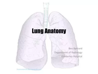
The Anatomy of Collaborative Staging:Lung Presentation developed by April Fritz, RHIT, CTR april.fritz@nih.gov SEER Program National Cancer Institute
Lung Cancer • Estimated 12.7% of all 2004 cancer cases • Collaborative Stage fields • Tumor Size--special codes • Extension • TS/Ext Eval--non-standard mapping • Lymph Nodes • LN Eval--non-standard mapping • LN Pos--standard • LN Exam--standard • Mets at Dx • Mets Eval--standard • Site-specific factors 1-6--not used
Lung -- Tumor Size • Site-specific (not standard) table • Special codes • 000-990 Standard definitions • 991-995 Less than _ cm • Use if precise size not available • 996 Occult carcinoma (TX) • (malignant cells in bronchopulmonary • secretions; no tumor seen) • 997 Diffuse (entire lobe) (M1) • 998 Diffuse (entire lung) (M1)
Lung -- CS Extension -- Notes 1. Direct extension to other structures is M1. Sternum, skeletal muscle, skin of chest, contra- lateral lung or mainstem bronchus, separate tumor nodules in different lobe 2. If resection done, assume tumor is > 2 cm from carina. 3. Assume opposite lung is not involved unless mentioned on x-ray/imaging. 4. Do not include bronchopneumonia with atelectasis in code 40 or 55. 5. Involved pulmonary artery/vein must be inside pericardium to be coded as 70.
Lung -- CS Extension -- Notes 6. Pleural effusion a. ignore negative pleural effusion (not 72) b. assume negative if resection performed c. ignore if clinical judgement says effusion is not related to tumor 7. Vocal cord paralysis--SVC obstruction-- compression of trachea/esophagus a. use Extension code 70 unless tumor is peripheral b. use LN code 20 if tumor is peripheral • Code an extension where it appears. • Computer algorithm will correctly assign the stage
B C E F D A Lung -- CS Extension -- 00-11, 23-30 Code 00 Very rare Code 10 Confined to lung; needs size Code 11 Superficial bronchus only (D) Code 23 Starts in hilus (E); needs size Code 25 Starts in carina (F); needs size Code 30 Localized, NOS CS Extension Code 10 Tumor surrounded by lung (A) or visceral pleura (B); no invasion more proximal than a lobar bronchus (C)
C A >2 cm B Lung -- CS Extension -- 20-21, 45 CS Extension Codes 20 In main bronchus > 2 cm from carina (A) 21 Involving mainstem bronchus, distance not stated (B) 45 Invading visceral pleura (C)
B A Lung -- CS Extension -- 40 Extension Code 40 Tumor associated with atelectasis (A) or obstructive pneumonitis (B) that extends to the hilar region but does not involve entire lung; no pleural effusion
2 cm B A Lung -- CS Extension -- 50-55 Extension Codes 50 Tumor in main bronchus < 2 cm from carina without involving carina (A); also 52 and 53 55 Atelectasis or obstructive pneumonitis of entire lung (B)
Superior sulcus Trachea Clavicle B E A D C Ribs D Pleura Pleural space Pericardium Diaphragm Lung -- CS Extension -- 56-61 Extension Codes Direct extension to: 56 Parietal pericardium (A) 59 Phrenic nerve (not shown) 60 Brachial plexus from superior sulcus (B); Pancoast tumor 60 Chest wall (C) 60 Diaphragm (D) 60 Parietal pleura (E) 61 Upper brachial plexus (not shown, similar to B)
C D E D B A Lung -- CS Extension -- 70, 71, 73, 75 Direct invasion of any of the following: 70 Mediastinum (A) 71 Heart, visceral pericardium (B) 70 Trachea (C) 70 Great vessels (D) 70 Carina (E) Not shown: 70 Esophagus (behind trachea) 70 Nerves 73 Adjacent rib 75 Vertebral body (posterior to lung)
70 Superior vena cava 70 Main pulmonary artery 70 R and L pulmonary artery trunks* 70 R and L superior pulmonary veins* 70 R and L inferior pulmonary veins* 74 Aorta 77 Inferior vena cava * intrapericardial segments Lung -- CS Extension -- Great Vessels
C A B Pleura Pleural effusion (malignant or NOS) Pleural space Lung -- CS Extension -- 65, 72 Extension Codes 65 Separate tumor nodules in same lobe (A) 72 Any tumor with malignant pleural effusion (B) 76 Discontinuous pleural tumor foci (C) 79 Pericardial effusion (not shown)
Lung -- CS Extension Discontinuous Nodules • Discontinuous tumor foci in ipsilateral parietal and visceral pleura from direct pleural invasion by primary tumor: Extension code 76 • Discontinuous tumors outside the parietal pleura in chest wall or diaphragm: Mets at Dx code 40
Lung -- CS Extension Remaining Extension Codes 80 Further contiguous extension (other than structures specified in Mets at Dx) 95 No evidence of primary tumor (T0) 98 Occult carcinoma (malignant cells in sputum or bronchial washings but lesion not seen) 99 Unknown extension; not assessed; not documented
Lung -- CS TS/Ext Eval • Non-standard mapping for TS/Ext Eval • Code 1 maps to pathologic • Includes endoscopic biopsies, FNA, surgical observation • Linked to CS Extension and Tumor Size • Document farthest extension clinically or pathologically • May not be highest eval code • Document information most useful for staging • Tumor size where appropriate • Extension where appropriate
Lung -- Coding CS TS/Ext Eval • Example 1 • Lung cancer, CXR shows 4 cm mass in medial RUL. Mediastinoscopy and FNA bx shows direct tumor extension through pleura into anterior mediastinum. Patient referred for radiation therapy. Codes: Tumor size 040 clinical (CXR) Extension 70 mediastinal extension TS/Ext Eval 1 endoscopic, FNA. Extension determines the mapping (pT4).
Lung -- Coding CS TS/Ext Eval • Example 2 • Lung mass, CXR shows 3.5 cm mass in RML. FNA shows squamous carcinoma. Resected specimen shows that tumor is surrounded by normal tissue but tumor size is actually 2.8 cm. Codes: Tumor size 028 path specimen Extension 10 confined to one lung TS/Ext Eval 3 surgical resection, no pre-op treatment Tumor size from path report determines the mapping (pT1).
Lung -- Coding CS TS/Ext Eval • Example 3 • Lung 5 cm RLL mass on CXR. CT scan shows pleural effusion on right. FNA of mass shows small cell carcinoma. Codes: Tumor size 050 path specimen Extension 72 pleural effusion (NOS) TS/Ext Eval 0 clinical (CT scan) Clinical findings document farther extension than tissue findings.
Lung -- CS Lymph Nodes -- Notes 1. Code only regional nodes in this field. 2. ‘Mass,’ ‘adenopathy’ or ‘enlargement’ of any nodes in code 20 are assumed to be involved. 3. Assume nodes are negative if stated as ‘No evidence of spread’ or ‘remaining exam negative’ and no other comment about nodes. 4. Vocal cord paralysis--SVC obstruction-- compression of trachea/esophagus a. use Extension code 70 unless tumor is peripheral b. use LN code 20 if tumor is peripheral and no statement of direct extension from a primary tumor
60 20 60 10 60 20 Lung -- CS Lymph Nodes Lymph Nodes 10 Same side Hilar, bronchial, peribronchial, intrapulmonary (LN stations 10-14) 20 Same side Subcarinal, mediastinal, others (LN stations 1-9) 50 Regional LN, NOS 60 Contralateral Mediastinal, hilar any scalene, any supraclavicular 80 Lymph nodes, NOS 99 Unknown, undocumented Adapted from R S Snell: Clinical Anatomy for Medical Students, 5th ed. 1995.
Lung -- CS Lymph Nodes Lymph Node Stations • Based on surgical landmarks • Not the same as LN codes • Station CS LN • 1-9 ipsilat 20 • 1-9 contralat 60 • 10-14 ipsilat 10 • 10-14 contralat 60 Source: Workbook for Staging of Cancer, 2nd ed., pages 110-111
Lung -- CS Reg Nodes Eval • Non-standard mapping for Reg Nodes Eval • Code 1 maps to pathologic • Includes endoscopic biopsies, FNA, surgical observation • Document farthest extension clinically or pathologically • May not be highest eval code • Document information most useful for staging
Lung -- Coding Reg Nodes Eval • Example 1 • Lung cancer, CXR shows 4 cm mass in right hilum. Mediastinoscopy and FNA bx of left hilar nodes shows poorly differentiated adenocarcinoma. Patient referred for radiation therapy. Codes: CS Lymph Nodes 60 Contralateral hilar Reg Nodes Eval 1 FNA lymph nodes Farthest involved lymph nodes confirmed by FNA (pN3). No need for complete resection of nodes.
Lung -- Coding Reg Nodes Eval • Example 2 • Lung mass, CXR shows left hilar mass, likely involved LN. FNA shows squamous carcinoma. Physical examination indicates hard left supraclavicular lymph node. Pt referred to medical oncologist. Codes: CS Lymph Nodes 60 Ipsilat. supraclav LN Reg Nodes Eval 0 Clinical Although hilar nodes (code 10) are proven by bx, clinical exam documents farther extension (cN3).
Lung -- CS Mets at Dx -- Notes • 1. For Mets at Dx, M0 or M1 is decided on • the basis of Tumor Size. • If Tumor Size is 998 (diffuse), Mets at Dx is M1 • For any other Tumor Size, Mets at Dx is M0
C A separate contralateral tumor nodule Primary tumor B separate ipsilateral tumor nodule D liver metastasis Lung -- CS Mets at Dx CS Mets at Dx Codes 10 Distant lymph nodes (A) Separate tumor nodules in a different lobe: 35 Ipsilateral (B); 39 Contralateral (C) 40 Distant metastasis (D) Not shown: 37 Extension to sternum, skeletal muscle, skin of chest 39 Extension to contralat lung, mainstem bronch. 50 (10 + 40)