Download
1 / 19
270 likes | 1.16k Views
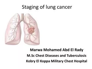
Lung Cancer: Staging And Prognosis. Hrach Ike Kasaryan. INTRODUCTION. Lung Cancer is the leading malignant cause of death in both men and women Ahead of Breast in women Ahead of Prostate in Men

E N D
Lung Cancer:Staging And Prognosis Hrach Ike Kasaryan
INTRODUCTION • Lung Cancer is the leading malignant cause of death in both men and women • Ahead of Breast in women • Ahead of Prostate in Men • It is important to have a good base of knowledge in internal medicine about how to stage Lung Cancers and what type of prognosis each stage carries.
Introduction • After discovering a patient has Lung Cancer, it is very important to stage the patient appropriately • There is a well established relationship between the ANATOMIC extent of the the disease and SURVIVAL DURATION • This is why staging a patient determines both prognosis and therapy
T/N/M system • The International System for Staging Lung Cancer is based on the TNM system • T-primary tumor • N-regional lymph nodes • M-distant metastasis • The system is applicable for the four major cell types of lung cancer: adenocarcinoma, squamous cell carcinoma, large cell carcinoma, and small cell carcinoma.
T/N/M system • This is based on all diagnostic and evaluative information obtained before the start of treatment, including the results of invasive procedures, such as bronchoscopy, needle biopsy, mediastinoscopy, and diagnostic thoracoscopy • The importance of Staging cannot be overemphasized because this information is to serve as a guide for treatment planning
Patients with Non-Small Cell & Small Cell Survival Curves based on staging
T- Primary Tumor • TX -Primary tumor cannot be assessed or tumor proved by the presence of malignant cells in sputum or bronchial washings but not visualized by imaging or bronchoscopy • TO - No evidence of primary tumor • Tis - Carcinoma in situ • T1 Tumor 3 cm or less in greatest dimension, surrounded by lung or visceral pleura, without bronchoscopic evidence of invasion more proximal than the lobar bronchus* (i.e., not in the main bronchus)
T – Primary Tumor • T2 - Tumor with any of the following features of size or extent: • More than 3 cm in greatest dimension • Involves main bronchus, 2 cm or more distal to the carina • Invades the visceral pleura • Associated with atelectasis or obstructive pneumonitis that extends to the hilar region but does not involve the entire lung
T- Primary Tumor • T3 - Tumor of any size that directly invades any of the following: • chest wall (including superior sulcus tumors), diaphragm, mediastinal pleura, or parietal pericardium; • or tumor in the main bronchus less than 2 cm distal to the carina but without involvement of the canna; • or associated atelectasis or obstructive pneumonitis of the entire lung
T- Primary Tumor • T4 - Tumor of any size that invades any of the following: • mediastinum, heart, great vessels, trachea, esophagus, vertebral body, or caring; • or tumor with a malignant pleural or pericardial effusion°; • or with satellite tumor nodule(s) within the ipsilateral primary tumor lobe of the lung
N- Regional Lymph Nodes • NX - Regional lymph nodes cannot be assessed • NO - No regional lymph node metastasis • N1 - Metastasis to ipsilateral peribronchial and/or ipsilateral hilar lymph nodes and intrapulmonary nodes involved by direct extension of the primary tumor • N2 - Metastasis to ipsilateral mediastinal and/or subcarinal lymph node(s) • N3 - Metastasis to contralateral mediastinal, contralateral hilar, ipsilateral or contralateral scalene, or supraclavicular lymph node(s)
M – Distant Metastasis • MX - Presence of distant metastasis cannot be assessed • MO - No Distant metastasis • M1 - Distant metastasis present
What is the Stage? • Patient has a 1cm Squamous Cell Carcinoma in the Right Mainstem Bronchus on bronchoscopy, No metastasis is seen on CAT SCAN and Lymph nodes are negative on Throcoscopy. • T2 N0 M0
What is the Stage? • Patient has on CAT SCAN a 2cm mass in the middle of the Left Lower Lobe limited to terminal bronchioles on Bronchoscopy with no pleural invasion, with no extension seen, Ipsilateral Mediastinal Nodes positive and Liver Mets. • T1 N2 M1
What is the Stage? • Patient on Cat SCAN with a 1cm Nodule in the Right Apex, isolated with no extension, FNA shows Adenocarcinoma, and on RU-Lobectomy all nodes are negative with no sign of metastasis on CAT Scan • T1 N0 M0