Download
1 / 43
450 likes | 617 Views
Collaborative Staging System. Part II Data Elements And Codes Stuart Herna, CTR. Data Elements. CS Tumor Size CS Extension CS Tumor Size/Ext Eval CS Lymph Nodes CS Reg Nodes Eval Regional LN Examined Regional LN Positive CS Mets At Dx CS Mets Eval CS Site Specific Factors 1-6.

E N D
Collaborative Staging System Part II Data Elements And Codes Stuart Herna, CTR
Data Elements • CS Tumor Size • CS Extension • CS Tumor Size/Ext Eval • CS Lymph Nodes • CS Reg Nodes Eval • Regional LN Examined • Regional LN Positive • CS Mets At Dx • CS Mets Eval • CS Site Specific Factors 1-6
Collecting the T Equivalent Computer derives TNM Registrar records cT1 pT2 yT3 cTX etc. CS Tumor Size (3 digits) T CS Extension (2 digits) CS TS/Ext Eval (1 digit)
CS Tumor Size CS Tumor Size records the largest lateral dimension or diameter of the primary tumor in millimeters, including melanoma • If no pre-operative treatment was performed, always code Pathologic Tumor Size • If pre-operative treatment was performed, code pre-treatment Clinical Tumor Size • Record the size of the invasive component of the tumor • Microscopic residual tumor does not affect Tumor Size
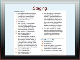
Coding CS Tumor Size (Lung) T0 T1/T2 T2 T1 T1/T2 TX T4 M1 TX 000 No mass/tumor found 001 - 988 001 - 988 millimeters (code exact size in millimeters) 989 989 millimeters or larger 990 Microscopic focus or foci only, no size of focus given 991 Described as less than 1 cm … 995 Described as less than 5 cm 996 Malignant cells present in bronchopulmonary secretions, but no tumor seen radiographically or during bronchoscopy; "occult" carcinoma 997 Diffuse (entire lobe) 998 Diffuse (entire lung or NOS) 999 Unknown; size not stated Not documented in patient record
CS Extension • CS Extension identifies contiguous growth of the primary tumor within the organ of origin or its direct extension into surrounding tissues (except Corpus Uteri and Ovary) • Always code the farthest extension of the primary tumor • If no neo-adjuvant treatment was performed, use the information in the path report to determine extension • If neo-adjuvant treatment was performed, use pre- treatment clinical information to determine extension
Coding CS Extension (Lung) 00 In situ; noninvasive; intraepithelial 10 Tumor confined to one lung; NO extension or conditions in 20-80 (EXCLUDES primary in main stem bronchus) (EXCLUDES superficial tumor as described in code 11) 11 Superficial tumor of any size with invasive component limited to bronchial wall, with or without proximal extension to the main stem bronchus 20 Extension from other parts of lung to main stem bronchus, NOS (EXCLUDES superficial tumor as described in code 11) Tumor involving main stem bronchus > 2 cm from carina (primary in lung or main stem bronchus) 21 Tumor involving main stem bronchus, NOS (distance from carina not stated, no surgery) Tis T1/T2* *based on size T1 T2 T2
Coding CS Extension (Lung) T1/T2 T1/T2 T1 T2 T2 T3 T3 T3 23 Tumor confined to hilus 25 Tumor confined to the carina 30 Localized, NOS 40 Atelectasis/obstructive pneumonitis that extends to the hilar region but does not involve the entire lung (or NOS) WITHOUT pleural effusion 45 Extension to (without pleural effusion): Pleura, visceral or NOS Pulmonary ligament 50 Tumor of/involving main stem bronchus < 2.0 cm from carina 52 (40) + (50) 53 (45) + (50)
Coding CS Extension (Lung) 55 Atelectasis/obstructive pneumonitis involving entire lung 56 Parietal pericardium or pericardium, NOS 59 Invasion of phrenic nerve 60 Extension to: (discontinuous ext.--see Mets at Dx) Brachial plexus (inferior branches or NOS) from superior sulcus Chest (thoracic) wall Diaphragm Pancoast tumor (superior sulcus syndrome) Parietal pleura 61 Superior sulcus tumor with encasement of subclavian vessels or uniquivocal involvement of superior branches of brachial plexus 65 Multiple masses/separate tumor nodule(s) in the SAME lobes; “Satellite nodules” in same lobe T3T3 T3 T3 T4 T4
Coding CS Extension (Lung) T4 T4 T4 T3 70 Blood vessel(s) (major): Azygos vein, Pulmonary artery or vein, Superior vena cava (SVC syndrome) Carina from lung/mainstem bronchus Compression of esophagus or trachea not specified as direct extension Esophagus Mediastinum, extrapulmonary or NOS Nerve(s): Cervical sympathetic (Horner's syndrome), Phrenic, Recurrent laryngeal (vocal cord paralysis), Vagus Trachea 71 Heart Visceral pericardium 72 Malignant pleural effusion Pleural effusion, NOS 73 Adjacent rib
Coding CS Extension (Lung) 74 Aorta 75 Vertebra(e) Neural foramina 76 Pleural tumor foci separate from direct pleural invasion 77 Inferior vena cava 79 Pericardial effusion, NOS; malignant pericardial effusion 80 Further contiguous extension (see also Mets at Dx) 95 No evidence of primary tumor 98 Tumor proven by presence of malignant cells in sputum or bronchial washings but not visualized by imaging or bronchoscopy; "occult" carcinoma 99 Unknown extension; Primary tumor cannot be assessed; Not documented in patient record T4 T4 T4 T4 T4 T4 T0 TX TX
CS Tumor Size/Ext Eval • CS Tumor Size/Ext Eval records how the codes for “CS Tumor Size” and “CS Extension” were determined based upon the methods employed (PE, Radiographic, Surgery, Autopsy, Etc.) • Select the code that documents the report or procedure from which the farthest tumor extension or size of the primary tumor was determined
Coding CS Tumor Size/Ext Eval (Lung) c p p p 0 PE, imaging, other non-invasive clinical evidence. No surgery; no autopsy 1 Endoscopy, dx biopsy, incl. FNA biopsy, or other invasive techniques including surgical observation without biopsy. No surgery, no autopsy 2 No surgery; based on autopsy (tumor suspected or dx’d prior to autopsy) 3 Surgical resection, NO pre-surgical systemic tx or RT OR Surgical resection, Unknown if pre-surgical systemic tx or RT. Evidence acquired before tx, supplemented by information obtained from surgery and pathologic examination of the resected specimen.
Coding CS Tumor Size/Ext Eval(Lung) c y a c 5 Surgical resection with pre-surgical systemic tx or RT; TS/Ext based on clinical evidence 6 Surgical resection with pre-surgical systemic tx or RT; TS/Ext based on pathologic evidence 8 Autopsy only (tumor unsuspected or undiagnosed prior to autopsy) 9 Unknown if surgical resection done; Not assessed; cannot be assessed; Unknown if assessed; Not documented in patient record
Collecting the N Equivalent Computer derives TNM Registrar records CS Lymph Nodes (2 digits) cN0 pN1 yN0 cNX etc. N CS Reg Nodes Eval (1 digit) Regional LN Positive (2 digits) Regional LN Examined (2 digits)
CS Lymph Nodes CS Lymph Nodes identifies the regional lymph nodes involved with cancer at the time of diagnosis • Record the RLN chain farthest from the primary site that is involved with cancer, either Clinically or Pathologically • For inaccessible sites, record RLN’s as negative rather than unknown if no mention is made of RLN involvement and patient receives usual standard cancer directed treatment • If patient receives neoadjuvant treatment, code the farthest involved RLN’s based upon information prior to treatment • If RLN’s are more involved after neoadjuvant treatment, code the most extensive RLN involvement
Coding CS Lymph Nodes (Lung) N0 N1 N2 00 None; no regional lymph node involvement 10 Regional lymph nodes (LN), ipsilateral: Bronchial; Hilar (bronchopulmonary) (proximal lobar) (pulmonary root); Intrapulmonary: Interlobar, Lobar, Segmental, Subsegmental, Peri/parabronchial 20 Regional LN, ipsilateral: Aortic [above diaphragm], NOS--Peri/para-aortic, NOS (Ascending aorta {phrenic}, Subaortic {aortico-pulmonary window}); Carinal (tracheobronchial) (tracheal bifurcation); Mediastinal, NOS--Anterior, Posterior (tracheo- esophageal); Pericardial; Peri/paraesophageal; Peri/paratracheal, NOS--Azygos (lower peritracheal), Pre- and retrotracheal, NOS (Precarinal); Pulmonary ligament; Subcardial; Subcarinal
Coding CS Lymph Nodes (Lung) N1 N3 N1 NX 50 Regional Lymph Nodes, NOS 60 Contralateral/bilateral hilar (bronchopulmonary) (proximal lobar) (pulmonary root); Contra- lateral/bilateral mediastinal; Scalene (inferior deep cervical), ipsilateral or contralateral; Supraclavicular (transverse cervical), ipsilateral or contralateral 80 Lymph nodes, NOS 99 Unknown; not stated Regional lymph nodes cannot be assessed Not documented in patient record
CS Reg Nodes Eval • CS Reg Nodes Eval records how the code for “CS Lymph Nodes” was determined based upon the diagnostic methods employed • Select the code that indicates the report or procedure from which information about the farthest involved RLN’s was obtained
Coding CS Reg Nodes Eval (Lung) c p p p 0 PE, imaging, other non-invasive clinical evidence. No LN removed; no autopsy 1 Endoscopy, dx biopsy, incl. FNA biopsy, or other invasive techniques including surgical observation without biopsy. No LN removed, no autopsy 2 No LN removed; based on autopsy (tumor suspected or dx’d prior to autopsy) 3 LN removed; NO pre-surgical systemic tx or RT OR LN removed, unk. if pre-surgical systemic tx or RT
Coding CS Reg Nodes Eval (Lung) c y a c 5 LN removed with pre-surgical systemic tx or RT; LN based on clinical evidence 6 LN removed with pre-surgical systemic tx or RT; LN based on pathologic evidence 8 Autopsy only (tumor not suspected or dx’d prior to autopsy) 9 Unknown if surgical resection done; Not assessed; cannot be assessed; Unknown if assessed; Not documented in patient record
Regional Lymph Nodes Positive • Regional LN Positive records the exact number of RLN’s examined by the pathologist and found to be positive • The fields Regional Lymph Nodes Positive and Reg Ln’s Examined are based upon pathologic information only
Coding Regional LN Positive p p p p p c c 00 All nodes examined negative. 01 - 89 1 - 89 nodes positive (code exact number of nodes positive) 90 90 or more nodes positive 95 Positive aspiration of lymph node(s) 97 Positive nodes - number unspecified 98 No nodes examined 99 Unknown if nodes are positive; not applicable; Not documented in record
Regional LN Examined • Regional LN Examined records the total number of RLN’s that were removed and examined by the pathologist • The number of RLN’s examined is cumulative from all procedures that removed RLN’s through the completion of surgery in the first course of treatment
Coding Regional LN Examined 00 No nodes examined. 01 - 89 1 - 89 nodes examined (code exact number of nodes examined) 90 90 or more nodes examined 95 No regional nodes removed but aspiration of regional nodes performed 96 LN “sampling,” number unk/not stated 97 LN “dissection,” number unk/not stated 98 LN removed, number unk/not stated; procedure type not stated; nodes examined, number unk 99 Unknown if nodes were examined; not applicable or negative; Not documented in patient record
Collecting the M Equivalent Computer derives TNM Registrar records cM0 pM1a yM0 cMX etc. CS Mets at Dx (2 digits) M CS Mets Eval (1 digit)
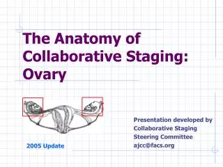
CS Mets At Dx • CS Mets At Dx identifies the distant site or sites of metastatic involvement at the time of diagnosis including distant lymph nodes • Assign the highest code for CS Mets At Dx whether determined clinically or pathologically and whether or not the patient had any neoadjuvant treatment
Coding CS Mets at Dx (Lung) 00 No distant metastases 10 Distant lymph node(s) 35 Separate tumor nodule(s) in different lobe, same lung 37 Extension to: Sternum, Skeletal muscle, Skin of chest 39 Extension to: Contralateral lung, Contralateral main stem bronchus; Separate tumor nodule(s) in contralateral lung 40 Distant metastases (other than code 10), except those in 35-39, including separate lesion in chest wall or diaphragm Distant metastasis, NOS; Carcinomatosis 50 (40) + (10); any of (35-39) + 10 99 Unknown; distant metastasis cannot be assessed; not stated in patient record M0 M1 M1 M1 M1 M1 M1 MX
CS Mets Eval • CS Mets Eval records how the code for data item “CS Mets at DX” was determined based upon the diagnostic methods employed • Select the CS Mets Eval code that documents the report or procedure from which the information was obtained about metastatic involvement farthest from the primary site • If the patient receives neoadjuvant treatment use the clinical status of metastatic involvement prior to treatment
Coding CS Mets Eval c c p p 0 PE, imaging, other non-invasive clinical evidence. No mets tissue examined; no autopsy 1 Endoscopy or other invasive techniques, including surgical observation without biopsy. No mets tissue examined, no autopsy 2 No mets tissue examined prior to death; based on autopsy (tumor suspected or dx’d prior to autopsy) 3 Mets tissue examined; NO pre-surgical systemic TX or RT OR Mets tissue examined, unk. if pre- surgical systemic TX or RT
Coding CS Mets Eval c y a c 5 Mets tissue examined with pre-surgical systemic TX or RT; Mets based on clinical evidence 6 Mets tissue examined with pre-surgical systemic TX or RT; Mets based on pathologic evidence 8 Autopsy only (tumor not suspected or dx’d prior to autopsy) 9 Unknown if surgical resection done; Not assessed; cannot be assessed; Unknown if assessed; Not documented in patient record
CS Site Specific Factors • CS Site-Specific Factors 1-6 identify additional information necessary to generate stage or prognostic factors that effect stage or survival including HIV status and tumor markers
Collecting Site-Specific Factors (SSF) • Replace existing “tumor marker” fields • Necessary for AJCC TNM changes (6th ed.) • Only used as needed by disease site
Example: SSF2 Melanoma Ulceration Note: Melanoma ulceration is the absence of an intact epidermis overlying the primary melanoma based upon histopathological examination. If no ulceration is documented in path report, code to 000. Code Description 000 No ulceration present adds ‘a’ to T1-T4 001 Ulceration present adds ‘b’ to T1-T4 999 Unknown; Not stated; Not documented in patient record
Example: LDH (SSF4 Melanoma; SSF3 Testis) LDH (Lactate Dehydrogenase) Code Description 000 Not done (SX) 002 Within normal limits (SO) 004 Range 1 (S1) < 1.5 x upper limit of normal for LDH assay 005 Range 2 (S2) 1.5 - 10 x upper limit of normal for LDH assay 006 Range 3 (S3) > 10 x upper limit of normal for LDH assay 008 Ordered, but results not in chart 999 Unknown; Not stated; Not documented in patient record Note: Per AJCC 6th ed. page 211, “An elevated serum LDH should only be used when there are two or more determinations obtained more than 24 hours apart, because an elevated serum LDH on a single determination can be falsely positive as a result of hemolysis or other factors unrelated to melanoma metastases.”
Collaborative Staging System Coding Practicum Lung
Example: Lung Cancer Case 1 6 months PTA: minimal cough; night sweats; 5-10 lb weight loss. 1½ PPD cigarette smoker x 30 years. PE: No lymphadenopathy or organomegaly. CXR: Solitary 3 cm. nodule in right upper lobe CT bone, brain and liver: within normal limits Bronchoscopy with biopsy: undiff large cell ca. Bronchoscopy brushings: positive for malignant cells Right upper lobectomy: 2.5 cm right upper lobe tumor; No gross hilar or mediastinal nodes noted. Pathology: Nodular 4 cm. mass showing poorly differentiated adenocarcinoma of right upper lobe, 0/12 hilar lymph nodes positive.
Example: Lung Case 1--Coded CS Tumor Size 040 4 cm per path report CS Extension 10 confined to one lung 3 resection, no pre-op (p) CS TS/Ext Eval CS Lymph Nodes 00 lymph nodes negative CS Reg Nodes Eval 3 resection, no pre-op (p) 00 per path report Reg LN Pos Reg LN Exam 12 per path report CS Mets at Dx 00 none 0 clinical (imaging) only (c) CS Mets Eval Site Specific Factors SSF1 - SSF6 888 not applicable
Example: Lung Case 1--Derived Derived Stage • TNM pT2 pN0 cM0 Stage IB • SS 1977 Localized SS 2000 Localized
Example: Lung Cancer Case 2 HX: Chest pain, productive cough, hoarseness with partial vocal cord paralysis. One PPD x 40 years. PE: Wheezing on expiration in both lungs. Otherwise no abnormal findings. CXR: 6 cm. RUL mass, incomplete atelectasis same lung. Pneumonitis and pleural effusion apparent. Scans of liver, brain: negative Bronchoscopy with biopsy: Right upper lobe mass noted with extension along lateral wall of main stem bronchus involving trachea. Washings and brushings positive for malignant cells. Scalene node biopsy (one node removed): metastatic SCC. Lung biopsy: Squamous cell carcinoma, poorly differentiated.
Example: Lung Case 2--Coded CS Tumor Size 060 6 cm per CXR 72 pleural effusion CS Extension 1 endoscopy CS TS/Ext Eval 60 scalene lymph nodes CS Lymph Nodes CS Reg Nodes Eval 3 LN bx established highest N Reg LN Pos 01 scalene node 01 scalene node Reg LN Exam CS Mets at Dx 00 none CS Mets Eval 0 clinical (imaging) only Site Specific Factors SSF1 - SSF6 888 not applicable
Example: Lung Case 2--Derived Derived Stage • TNM cT4 pN3 cM0 Stage IIIB • SS 1977 Distant SS 2000 Distant
CS Training Opportunities • Workshops • State and regional associations • Web-based training module • www.training.seer.cancer.gov • CS website • Presentations and exercises available • Algorithms, updates, additional information • www.cancerstaging.org/collab.html