Download
1 / 49
490 likes | 512 Views
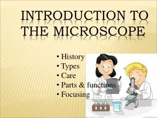
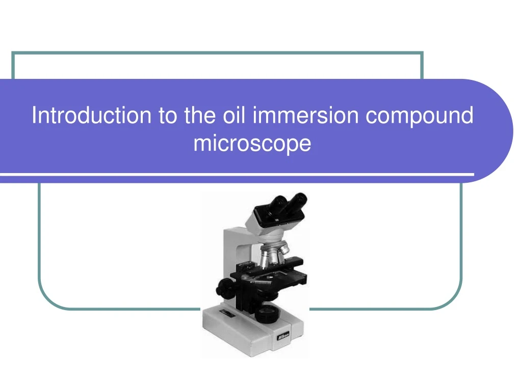
Introduction to the oil immersion compound microscope. Purpose of microscope. Microscopy is the technology of making very small things visible to the human eye. Used in the diagnosis of many diseases. Used for soil, insects and rocks. Used in pathological laboratories and medical.

E N D
Purpose of microscope • Microscopy is the technology of making very small things visible to the human eye. • Used in the diagnosis of many diseases. • Used for soil, insects and rocks. • Used in pathological laboratories and medical. • Industrial microscope : Used for metals and various kinds of measurement . • Educational microscope. • Used for research.
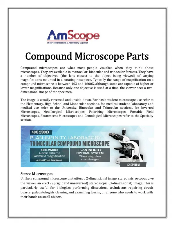
Purpose of microscope in Microbiology • Bacteria is one of the small microorganisms that can’t be seen with the naked eyes, most bacteria range in size between 0.5-2.0 micrometers (μm)so, there is a need to magnify the bacteria several times by using a microscope in order to see it. • There are different types of microscopes which are used in microbial life. • In this lab, you will become familiar with the use of the light compound microscope (particularly oil immersion microscopy) and will compare the relative size and shape of various microorganisms.
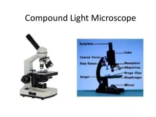
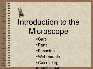
Microscope consists of 4 main parts: • frame work. • Adjustment system. • Magnification. • Lighting System.
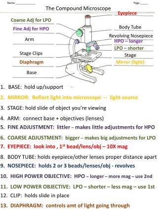
Frame work Its includes three parts: • Base • Arm • mechanical stage
Mechanical Stage • Stage- the location of the specimen to be viewed • Clips- utilized in holding the specimen in place
Stage Knobs control • Top knob: is to move the forward and backward. • Bottom knob: is to move the stage right and left.
Adjustment system It consists of the following parts: • Optical tube. • Coarse adjustment. • Fine adjustment.
Focus and Resolution Parts • Course-adjustment knob- is the larger of the two knobs. It is used in bringing the object into quick focus. • Fine-adjustment knob- is used for improving the clarity of the image, especially when viewing under high power.
Interpupillary adjustment To control the distance between the ocular lenses to adapt the distance between viewers eyes so, the eyepiece lenses will spread apart or get closer together to fit each individual.
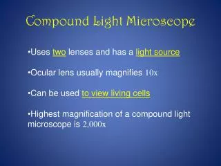
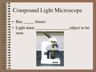
Magnification Microscope has two sets of lenses are : • Ocular lens (eyepiece). • Objetive lenes.
Microscope lenses: • Ocular lens or eyepiece is used for viewing. • Revolving nosepiece contains objective lenses that are used to magnify the image in combination with the ocular lens. • The objective of microscopy is not just to increase magnification, but to do so while retaining sufficient resolution.
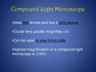
Important points around the light microscope: • Total magnification = • Eyepiece magnification X Objectives magnification. • The image seen by the eye through a compound microscope is termed the virtual image and is upside and reversed.
Important points around the light microscope: Total magnifigation: Powers of theeyepiece (10X) multiplied by objective lenses determine total magnification.
Important points around the light microscope: • The numerical aperture (NA): • Is a designation of the amount of light entering the objective from? • The microscopic field • NA = R sin • Where: • R is the refractive index of glass • is the angle made by one ray passing through • the edge of the lens with the other ray passing the center of the lens. • Since sin = AC/AP • Then NA depends on the radius of the lens.
Important points around the light microscope: 3. Resolving power: • Is the useful limit of magnification, it is the ability of microscope, at specific magnification to distinguish two separate objects situated close to one another and the ability of the lens to reveal fine details. • The smaller the distance between the two specific objects that can be distinguished apart, the greater the resolution power of the microscope. • Minimal distance between two objects = (0.612 X ) / NA • The larger NA, the smaller the resolvable distance and hence, the more efficient the resolution power.
Resolution • The objective of microscopy is not just to increase magnification, but to do so while retaining sufficient resolution. • Resolution: It is the ability of microscope, at specific magnification to distinguish two separate objects situated close to one another and the ability of the lens to reveal fine details. • The smaller the distance between the two specific objects that can be distinguished apart, the greater the resolution power of the microscope.
As magnification increased the resolution also increased. • To achieve high magnification with good resolution the objective lens must be small. • Shorter wavelengths of light provide greater resolution. • Under the best conditions, with violet light (wavelength = 0.4 um) and a numerical aperture of 1.4, the light microscope can theoretically achieve a limit of resolution of just under 0.2 pm.
A typical cell is 10-20 um Light microscope sees 0.2 um 1 mm=10-3 m 1 um=10-6 m 1 nm=10-9 m 1 A=Angstrom =10-10 m
Important points around the light microscope: • Depth of field: • Is the capacity of the objective lens to focus in different planes at the same time. • This is largely dependent on the NA. • Where the greater the NA, the smaller the depth of the field, it is possible to increase the depth of the field slightly by closing the iris diaphragm. Thus decrease the NA
Depth of focus: • thickness of the field; • as magnification increased , depth of focus is decreased. • Field of vision: • the surface area of view; • as magnification increased , the area of view decreased.
The mediumbetween the objective and the object is a factor that must be taken into consideration for the most effective use of the microscope lens. The low power objective 4x, 10x, and high dry objective 40x use air. When oil immersion lenses are employed, a drop of oil should be used, otherwise bending of the light waves occurs since oil has the same RI of glass, while air increase diffraction.
Refraction and Refractive Index • Refraction = bending. • Refractive index: measurement of the extent that the substance bend light. • Field of view > or equal 500X cause increase in diffraction due to • the differences in refractive index between glass and lenses (1.51) and air (1) which increase refraction, • and small lenses present in high magnification • so that oil is used in these lenses as it has refractive index similar to glass, lenses and specimen which equal 1.51 ,as a result the distortion will decrease.
Lighting System: Lighting system consists of modern microscopes of the following parts : • Power supply • Transformer • Variable resistance • light source • sub stage condenser • Aperture diaphragm
Lighting System: • The Condenser (fucous light through specimen ) • Lamp- typically a light source underneath the stage • Diaphragm- controls the amount of light allowed to pass through the specimen
How to use Microscope • Plug in microscope and turn on illuminator. Rotate nosepiece to lock 4X objective in place • Place smear on stage and center it under the 4X objective. • Using the course adjustment knob, move the objective lens to its lowest point. Look through the ocular and focus upward with the coarse adjustment until an image comes into view. • Rotate nosepiece to obtain the next objective lens 10x and repeat step 3. • Rotate nosepiece to obtain the next objective lens 40x . • Look through the ocular and focus upward with the fine adjustment until an image comes into view. • When using oil immersion lens put a drop of oil on the slide and Rotate nosepiece and repeat step 6.
With a binocular microscope, adjust oculars for both eyes! With a monocular microscope, keep both eyes open!
Factors that influence the quality and size of image: • Quality of Microscope and lenses. • Size of sample. • Type of sample. • Amount of light on the sample. • Quality of sample.
Microscope Storage • Proper storage of the microscope will prevent or reduce problems! • Optics and mechanisms of the microscope must be protected from: • Dust and dirt, • Fungus. • Store the microscope: • Under a protective cover, • In a low humidity environment. Always carry a microscope with one hand holding the arm and one hand under the base.
Microscope Cleaning Process • Cleaning the Eyepiece, • Cleaning the Objectives, • Cleaning the Microscope Stage, • Cleaning the Microscope Body, • Cleaning the Condenser.
Cleaning Solutions and Solvents • Soap solution for cleaning of body and stage. • Ether-Alcohol, Alcohol, or Lens Cleaner Solution for cleaning of lenses. • Refer to manufacturer’s guide for appropriate organic solvent.
Cleaning Materials • Lint-free cotton gauze pads • Lint-free cotton swabs • Lens paper • Commercial lens tissue for optics • Caution: Do not use paper towels or other rough paper products • Alternatives include: • Fine quality tissue paper • Muslin cloth • Silk
Microscope Cleaning Process • Cleaning the Eyepiece, • Cleaning the Objectives, • Cleaning the Microscope Stage, • Cleaning the Microscope Body, • Cleaning the Condenser.
Step 1: Cleaning the Eyepieces • Blow to remove dust before wiping lens. • Clean the eyepieces with a cotton swab moistened with lens cleaning solution. • Clean in a circular motion inside out.
Step 2: Cleaning the Eyepiece • Wipe the eyepieces dry with lens paper. • Repeat cleaning and drying if required.
Step 3:Cleaning the Objectives • Objectives are cleaned while attached to microscope • Moisten the lens paper with the cleaning solution • Wipe gently the objective in circular motion from inside out • Wipe with dry tissue or lens cleaning paper . • Be sure to clean the oil immersion objective after use. • Objectives should never be removed from the nosepiece.
Step 4: Cleaning the Microscope Stage • Wipe the microscope stage using the cleaning solution on a soft cloth. • Thoroughly dry the stage. • Repeat above steps, if required.
Step 6: Cleaning the Condenser • Unplug the microscope from power source. • Clean the condenser lens and auxiliary lens using lint-free cotton swabs moistened with lens cleaning solution. • Wipe with dry swabs.
Step 5: Cleaning the Microscope Body • Unplug the microscope from power source. • Moisten the cotton pad with a mild cleaning agent . • Wipe the microscope body to remove dust, dirt, and oil. • Repeat steps 1–3, if required.