Download
1 / 13
210 likes | 522 Views
The Compound Light Microscope. Parts of the Light Microscope. Stage – part of the microscope that holds the specimen for viewing Light source – light bulb located beneath the stage

E N D
The Compound Light Microscope Parts of the Light Microscope
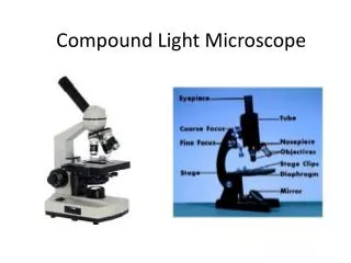
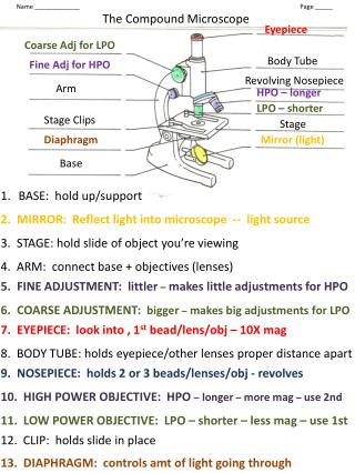
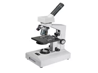
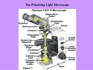
Stage – part of the microscope that holds the specimen for viewing • Light source – light bulb located beneath the stage • Adjustable diaphragm – located beneath the stage and is used to regulate the amount of light that passes through • The body tube contains an ocular lens (eyepiece) and a nosepiece with several objective lenses. Each objective lens is used for a different magnification and is moved into place by revolving the nosepiece. The image is brought into focus by adjusting the coarse and fine focus knobs.
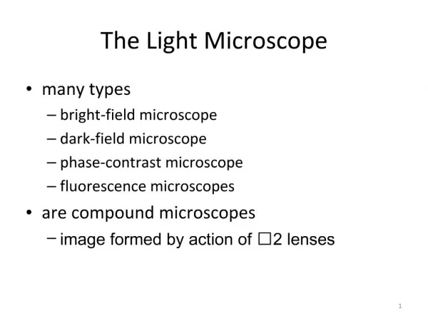
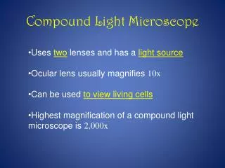
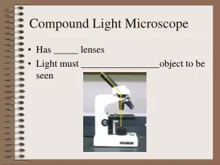
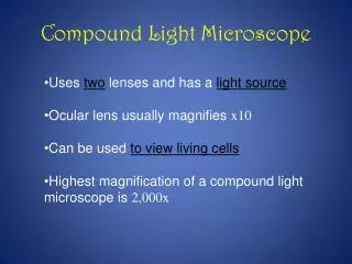
Compound light microscopes contain two lens systems, an objective and an ocular. The total magnification of an image is calculated by multiplying the magnification of the ocular by the magnification of the objective. The microscopes we will use each have a 10X ocular lens and three or four different objective lenses.
CAUTION - Never use cloth or paper products (paper towels, tissue paper, etc.) to clean the lenses. They will scratch the coating and decrease the resolving power of the lens. Use only lens paper. • Switch the microscope to the lowest magnification or raise the objectives from the stage before inserting a slide. This will prevent the objective lens from being accidentally scratched by the slide. • Place the slide to be viewed on the stage and center the specimen over the opening. • Begin with the low power objective lens.
Raise the stage all the way so that the slide is as close as possible to the objective lens. • Use the coarse adjustment knob to slowly raise the lens from the stage while viewing the image. Fine focusing is not needed when using the lowest magnification (scanning or 4X objective). If you are using any of the other objectives, it will be necessary to use the fine focus after using the coarse focus. • Adjust the diaphragm. This will need readjustment after changing to a different magnification.
To Increase the Magnification • The microscopes are parfocal, meaning that after you adjust the focus, the image will remain approximately in focus if you change the magnification. • Center the object before switching to a higher power objective. This will help you find the object after switching the objective. • Switch to the next highest power. It will be necessary to center the image again. The image should be approximately in focus but it will be necessary to use the fine focus. The coarse focus should not be needed after switching objectives. • Adjust the diaphragm. • This procedure is repeated each time you switch to a higher magnification
Placing a slide on the stage Begining with the low power lens Adjusting the magnification
Oil Emersion • The 100 X objective (1,000X total magnification) requires that a drop of immersion oil be placed between the slide and the lens. • After focusing the specimen under high power, rotate the high power objective out of the way and place a drop of immersion oil on the slide. Rotate the oil immersion objective into place so that it touches the oil. • Adjust the fine focus, condenser, and diaphragm as previously described. • After viewing with oil, the lens must be cleaned with fluid designated for this purpose. Remember, use lens paper only. Never use cloth, paper towels, or other paper products on coated optics.