Download
1 / 13
680 likes | 3.38k Views
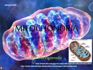
Tamboli Alija Z. Dept of Zoology, S.M.Joshi Collge , Hadapsar. F.Y.B.Sc. Mitochondria. Introduction. Mitochondria were first seen by kollicker in 1850 in muscles and called them ‘ sarcosomes ’ Flemming (1882) described these organelles as ‘filia’

E N D
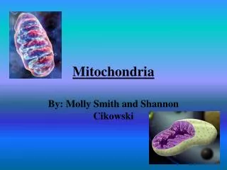
Tamboli Alija Z.Dept of Zoology, S.M.Joshi Collge , Hadapsar.F.Y.B.Sc Mitochondria
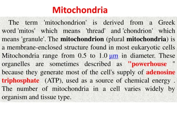
Introduction Mitochondria were first seen by kollicker in 1850 in muscles and called them ‘sarcosomes’ Flemming (1882) described these organelles as ‘filia’ Altmann (1890) observed these structures and named them ‘bioblasts’. Benda (1898)stained these organelles with crystalviolet and renamed them ‘mitochondria’ Michaelis (1900) used janus green B as a vital stain to observe mitochondria in living cells.
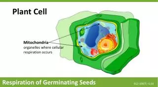
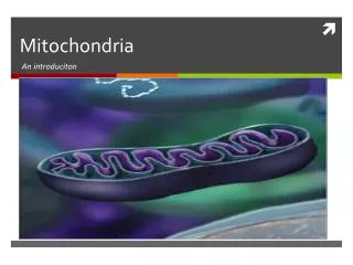
Morphology The shape of mitochondria is highly variable ranges from short rod shape to elongate filamentous form . The size of mitochondria is variable ,they generally measures about 0.5 to 2um in diameter. Mitochondria have an average length of 3 to 4um. The number of mitochondria varies from one cell type to another. Mitochondria are not found in prokaryotes.
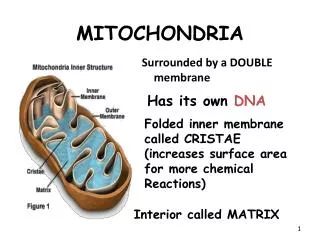
Ultra Structure A mitochondria is enclosed by a double membrane envelope composed of lipid and protein. The two membranes are separated by a narrow fluid –filled space called the outer compartment. The outer membrane is smooth, it is more permeable to small molecules, contains some enzymes but is poorer in proteins. The inner membrane surrounds a central cavity or matrix (inner compartment) filled with a fluid. Folds of inner wall of mitochondria are called cristae.
Inner Membrane Inner membrane is the site of the e- transport chain, across which the proton pump occurs and contains ATP synthase. Inner membrane is highly folded – called cristae – increasing the surface area on which the above reactions can take place
Mitochondrial Inner Membrane The inner mitochondrial membrane is compartmentalized into numerous cristae, which expand the surface area of the inner mitochondrial membrane, enhancing its ability to generate ATP. In typical liver mitochondria, for example, the surface area, including cristae, is about five times that of the outer membrane. Mitochondria of cells which have greater demand for ATP, such as muscle cells, contain more cristae than typical liver mitochondria.
Mitochondrial Outer Membrane The outer mitochondrial membrane, which encloses the entire organelle, has a protein-to-phospholipid ratio similar to the eukaryotic plasma membrane (about 1:1 by weight). It contains numerous integral proteins called porins, which contain a relatively large internal channel (about 2-3 nm) that is permeable to all molecules of 5000 daltons or less. Larger molecules, for example most proteins, can only traverse the outer membrane by active transport.
Mitochondria Double membrane creates 2 spaces Matrix – large internal space Intermembrane space – between the membranes Outer membrane Inner membrane
Chemical Composition Mitochondria consists of protein-70 percent & lipids -25 -30percent. Mitochondria contain 0.5percent of RNA & traces of DNA . Mitochondrial DNA comprises about 1 percent of total cell DNA Mitochondria contain enzymes for oxidation phosphorylation & electron transfer.
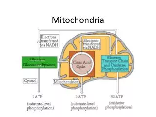
Mitochondria Produce most of a cells ATP – acetyl groups in the Kreb’s cycle producing CO2 and NADH. NADH donates the e- to the electron transport chain and becomes oxidized to NAD+ e- transfer promotes proton pump and ATP synthesis in process called oxidative phosphorylation Cells that require large amounts of energy such as the heart have large numbers of mitochondria
Mitochondria Contain their own copies of DNA and RNA along with transcription and translation system (ribosomes) Are able to regenerate themselves without the whole cell undergoing division Shape and size dependent on what the cell’s function is