Download
1 / 44
440 likes | 445 Views
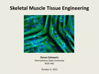
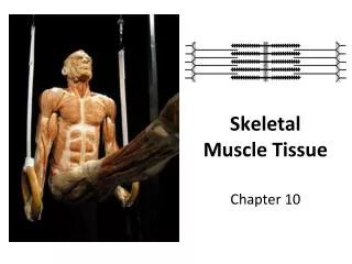
SKELETAL TISSUE AND PHYSIOLOGY. CARTILAGE. Connective tissue Fibers embedded in firm gel Avascular Chondrocytes lie in lacunae Nutrients delivered by diffusion. 3 TYPES. Fibrocartilage – menesci of knee and between vertebrae Elastic – external ear and epiglottis

E N D
CARTILAGE • Connective tissue • Fibers embedded in firm gel • Avascular • Chondrocytes lie in lacunae • Nutrients delivered by diffusion
3 TYPES • Fibrocartilage – menesci of knee and between vertebrae • Elastic – external ear and epiglottis • Hyaline – articular, chondral, laryngeal, tracheal and bronchial
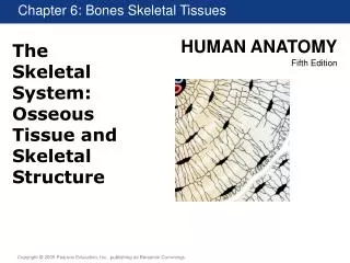
FUNCTIONS OF SKELETON • Support • Protection • Movement –bones and joints – levers • Mineral reservoir – Ca++, Phosphorus • Hemopoiesis – blood cell formation
TYPES OF BONES • Long – humerus, femur, ulna, radius, tibia, fibula • Short – carpals and tarsals • Flat – skull, ribs, patella, scapula • Irregular – vertebrae, facial bones, hyoid
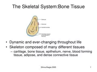
MACROSCOPIC STRUCTURE OF LONG BONES • Diaphysis – main shaft like portion; hollow, cylindrical, thick compact bone • Epiphysis(-es) – end of long bones – bulbous shape provides generous space for muscle attachments – spongy (cancellous) bone filled with yellow marrow except in proximal epiphyses of humerus and femur (red)
Articular cartilage – thin layer of hyaline cartilage that covers joint surfaces of epiphyses – shock absorber • Periosteum –dense, white fibrous membrane that covers bone everywhere but joint surfaces – Sharpey’s fibers penetrate bone – muscle fibers interlace with these providing a firm anchor – BVs from periosteum nourish bone –
- OSTEOBLASTS (bone building cells) compose inner periosteum 5. Medullary cavity – marrow containing cavity in diaphysis 6. Endosteum – membrane lining medullary cavity
SHORT, FLAT AND IRREGULAR BONES • Cancellous bone covered w/compact bone • Red marrow fills spaces in cancellous bone inside a few irregular and flat bones (vertebrae and sternum) • Needle aspiration – diagnostic tool
BONE TISSUE • Connective tissue – cells, fibers (collagen) and calcified matrix • More matrix than cells; lots of collagen • Strength of cast iron, 1/3 the mass
BONE MATRIX • Inorganic salts – APATITE (crystals of calcium and phosphate) gives bone its hardness – oriented to resist stress • Organic matrix – collagen and ground substance – amorphous mixture of protein and polysaccharides
MICROSCOPIC STRUCTURE OF BONE Compact bone composed of Haversian system • Lamellae – concentric cylinders of calcified matrix • Lacunae – small spaces containing tissue fluid in which osteocytes lie between lamellae • Canaliculi – small canals radiating from lacunae, connecting them to each other and the Haversian canal
Haversian canal – extends through center of each Haversian system – blood and lymph transport O2 and nutrients to bone cells - BVs from periosteum penetrate bone via Volkmann’s canals; arteries supply marrow Cancellous bone – NO Haversian systems; weblike arrangement of marrow filled spaces separated by trabeculae (thin processes of bone)
BONE MARKINGS • DEPRESSIONS AND OPENINGS • Fossa • Sinus • Foramen • Meatus • Sulcus
B. PROJECTIONS AND PROCESSES 1. Condyle 2. Head 3. Trochanter 4. Crest 5. Spinous process 6. Tuberosity 7. Tubercle
DEVELOPMENT OF BONE - OSTEOGENESIS 1. Intramembranous ossification • happens in connective tissue membrane • Includes broad flat bones of the skull • Membrane like layers of primitive connective tissue appear at the site of future bone • Layers supplied with blood – connective tissue cells arrange themselves around the blood vessel
These cells differentiate into osteoblasts • Osteoblasts deposit bony matrix – produce spongy bone • Osteoblasts become surrounded by bony matrix – in lacunae, osteocytes • Osteoblasts on inside of periosteum give rise to compact bone
2. Endochondral • Formed from hyaline cartilage model • Periosteum develops, enlarges, and forms subperiosteal collar • 1o ossification center develops as cartilage begins to calcify and BVs enter rapidly changing cartilage model at midpoint of diaphysis • Ossification proceeds from diaphysis to epiphysis • Bone grows in length
2o ossification center appears at epiphyses and growth proceeds from epiphysis to diaphysis • Until bone length growth is complete, a layer of cartilage (epiphyseal cartilage) remains between diaphysis and epiphysis • Epiphyseal cartilage thickens during growth periods • This cartilage ossifies – osteoblasts make organic bone matrix, matrix calcifies – bone grows longer
BONE GROWTH AND RESORPTION Bone growth – diameter • Osteoclasts • Osteoblasts • Ossification and resorption occur concurrently • In adult years, rate = • Childhood and adolescence, ossification > resorption • 35- 40 years, resorption > ossification
BONE FRACTURES AND REPAIRS Fracture – break in bone’s continuity • Simple – skin unbroken • Compound – skin broken • Alignment = reduction • Closed reduction = fracture set without opening skin • Open reduction – requires surgical incision • Osteomyelitis • Kids – greenstick fractures – bone cracked
HEALING OF FRACTURES • Dead bone – removed by osteoclasts or serves as framework for CALLUS (repair tissue) • Callus – periosteal and endosteal cells differentiate into chondroblasts and osteoblasts • Callus binds broken ends – callus tissue is eventually replaced by normal bone