Download
1 / 49
490 likes | 706 Views
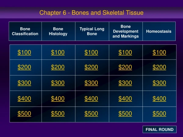
Bones and Skeletal Tissue. Bones and Cartilages of the Human Body. Figure 6.1. How are bones classified?. Axial skeleton – bones of the skull, vertebral column, and rib cage Appendicular skeleton – bones of the upper and lower limbs, shoulder, and hip. Long bones.

E N D
Bones and Cartilages of the Human Body Figure 6.1
How are bones classified? • Axial skeleton – bones of the skull, vertebral column, and rib cage • Appendicular skeleton – bones of the upper and lower limbs, shoulder, and hip
Long bones • Long bones – longer than they are wide (e.g., humerus) Figure 6.2a
Short bones • Short bones • Cube-shaped bones of the wrist and ankle • Bones that form within tendons (e.g., patella) Figure 6.2b
Flat bones • Flat bones – thin, flattened, and a bit curved (e.g., sternum, and most skull bones) Figure 6.2c
Irregular bones • Irregular bones – bones with complicated shapes (e.g., vertebrae and hip bones) Figure 6.2d
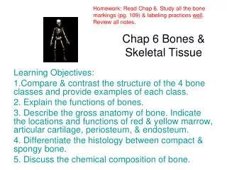
What are the functions of bones? • Support – form the framework that supports the body and cradles soft organs • Protection – provide a protective case for the brain, spinal cord, and vital organs • Movement – provide levers for muscles • Mineral storage – reservoir for minerals, especially calcium and phosphorus • Blood cell formation – hematopoiesis occurs within the marrow cavities of bones
What is the Gross Anatomy of Bones? • Compact bone – dense outer layer • Spongy bone – honeycomb of trabeculae (little beams) filled with red bone marrow
What is the structure of a long bone? • Diaphysis • Tubular shaft that forms the axis of long bones • Composed of compact bone that surrounds the medullary cavity • Yellow bone marrow (fat) is contained in the medullary cavity
Structure of Long Bone • Epiphyses • Expanded ends of long bones • Exterior is compact bone, and the interior is spongy bone • Joint surface is covered with articular (hyaline) cartilage • Epiphyseal line separates the diaphysis from the epiphyses
What are the bone membranes? • Periosteum – double-layered protective membrane • Outer fibrous layer is dense regular CT • Inner osteogenic layer is composed of osteoblasts and osteoclasts • Richly supplied with nerve fibers, blood, and lymphatic vessels, which enter the bone via nutrient foramina • Sharpey’s fibers: secures the underlying bone to the periosteum. They are tufts of collagen fibers. • Endosteum – delicate membrane covering internal surfaces of bone
What is the structure of short, irregular, and flat bones? • Thin plates of periosteum-covered compact bone on the outside with endosteum-covered spongy bone on the inside • Have no diaphysis or epiphyses • Contain bone marrow between the trabeculae Figure 6.4
Where is the location of hematopoietic tissue (Red Marrow)? • In infants • Found in the medullary cavity and all areas of spongy bone • In adults • Found in the middle of flat bones, and the head of the femur and humerus
What is microscopic structure of bone: compact bone? • Haversian system, or osteon – the structural unit of compact bone • Lamella – weight-bearing, column-like matrix tubes composed mainly of collagen • Haversian, or central canal – central channel containing blood vessels and nerves • Volkmann’s canals – channels lying at right angles to the central canal, connecting blood and nerve supply of the periosteum to that of the Haversian canal
Microscopic Structure of Bone: Compact Bone • Osteocytes – mature bone cells • Lacunae – small cavities in bone that contain osteocytes • Canaliculi – hairlike canals that connect lacunae to each other and the central canal
What is the chemical composition of bone? (Organic) • Osteoblasts – bone-forming cells • Osteocytes – mature bone cells • Osteoclasts – large cells that reabsorb or break down bone matrix
Chemical Composition of Bone: Inorganic • Hydroxyapatites, or mineral salts • Sixty-five percent of bone by mass • Mainly calcium phosphates • Responsible for bone hardness and its resistance to compression
What are the types of markings found on bones? • Bulges, depressions, and holes that serve as: • Sites of attachment for muscles, ligaments, and tendons • Joint surfaces • Conduits for blood vessels and nerves
Bone Markings: Projections – Sites of Muscle and Ligament Attachment • Tuberosity – rounded projection • Crest – narrow, prominent ridge of bone • Trochanter – large, blunt, irregular surface • Line – narrow ridge of bone
Bone Markings: Projections – Sites of Muscle and Ligament Attachment • Tubercle – small rounded projection • Epicondyle – raised area above a condyle • Spine – sharp, slender projection • Process – any bony prominence
Bone Markings: Projections That Help to Form Joints • Head – bony expansion carried on a narrow neck • Facet – smooth, nearly flat articular surface • Condyle – rounded articular projection • Ramus – armlike bar of bone
Bone Markings: Depressions and Openings • Meatus – canal-like passageway • Sinus – cavity within a bone • Fossa – shallow, basinlike depression • Groove – furrow • Fissure – narrow, slitlike opening • Foramen – round or oval opening through a bone
How do bones develop? • Osteogenesis and ossification – the process of bone tissue formation, which leads to: • The formation of the bony skeleton in embryos • Bone growth until early adulthood • Bone thickness, remodeling, and repair
Formation of the Bony Skeleton • Begins at week 8 of embryo development • Intramembranous ossification – bone develops from a fibrous membrane • Endochondral ossification – bone forms by replacing hyaline cartilage
Functional Zones in Long Bone Growth • Growth zone – cartilage cells undergo mitosis, pushing the epiphysis away from the diaphysis • Transformation zone – older cells enlarge, the matrix becomes calcified, cartilage cells die, and the matrix begins to deteriorate • Osteogenic zone – new bone formation occurs
Long Bone Growth and Remodeling • Growth in length – cartilage continually grows and is replaced by bone as shown • Remodeling – bone is reabsorbed and added by appositional growth as shown Figure 6.10
Tendon • Generally regular connective tissue • Musculo-skeletal connections • Muscle to bone • Muscle to muscle • Bone to bone Fig. 4.15f, M&M Frolich, Human Anatomy, Mechanics of Movement
Tendons • Tendons are structures that connect bone to muscle and are made up of tendon tissue • Can have various shapes • Typical is cord-like tendon of biceps • Sheeths are common--”aponeuroses” e.g. acromiotrapezius origin from thoracic vertebral spines Fig. 10.3, M&M Frolich, Human Anatomy, Mechanics of Movement
Ligaments • Ligaments connect bone-to-bone or reinforce joints--they are made up of tendinous tissue as well • E.g. knee ligaments Fig. 9.12, M&M Frolich, Human Anatomy, Mechanics of Movement
Synovial Joint Shape Types • Plane joints--intercarpal joints • Hinge joints--elbow,ankle, interj-phalangeal • Pivot joints--radio-ulnar joint • Condyloid joints (egg into oval)--metacarpo-phalangeal • Saddle joints--carpo-metacarpal joint of thumb • Ball-and-socket--hip, shoulder The type of joint, in part, determines the range and direction of movement Frolich, Human Anatomy, Mechanics of Movement
Synovial Joints • Ball-and-Socket: The ball-shaped end of one bone fits into a cup shaped socket on the other bone allowing the widest range of motion including rotation. Examples include the shoulder and hip. • Condyloid: Oval shaped condyle fits into elliptical cavity of another allowing angular motion but not rotation. This occurs between the metacarpals (bones in the palm of the hand) and phalanges (fingers) and between the metatarsals (foot bones excluding heel) and phalanges (toes).
Saddle: This type of joint occurs when the touching surfaces of two bones have both concave and convex regions with the shapes of the two bones complementing one other and allowing a wide range of movement. The only saddle joint in the body is in the thumb. • Pivot: Rounded or conical surfaces of one bone fit into a ring of one or tendon allowing rotation. An example is the joint between the axis and atlas in the neck.
Synovial Joints • Hinge: A convex projection on one bone fits into a concave depression in another permitting only flexion and extension as in the elbow joints. • Gliding: Flat or slightly flat surfaces move against each other allowing sliding or twisting without any circular movement. This happens in the carpals in the wrist and the tarsals in the ankle.