Download
1 / 5

E N D
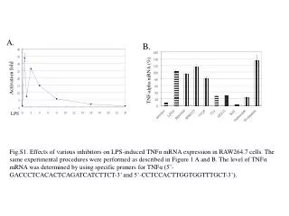
Fig.S1. Effects of various inhibitors on LPS-induced TNFα mRNA expression in RAW264.7 cells. The same experimental procedures were performed as described in Figure 1 A and B. The level of TNFα mRNA was determined by using specific primers for TNFα (5’-GACCCTCACACTCAGATCATCTTCT-3’ and 5’-CCTCCACTTGGTGGTTTGCT-3’).
Fig.S2. The inhibitory effect of 1 mM of SB203580 on LPS-induced TTP mRNA expression. RAW264.7 cells were pretreated with 1 mM of SB203580 for 30 min, and then followed by treated with LPS for 30 min or 2 h. The total RNAs were isolated for real-time PCR analysis by using specific primers for TTP.
Fig.S3 Induction of TTP expression by recombinant TNFα treatment in NIH3T3 cells.NIH3T3 cells were pretreated or untreated with 20 mM BAY for 30 min followed by treated with 10 ng/ml recombinant TNFα for 0, 15 min, 30 min and 60 min and the whole cell extracts were isolated for Western blotting analysis by using anti-TTP antibody. Anti-α-tubulin were used as internal control. BAY: - - - - + TNF a(min): 0 15 30 60 30 TTP tubulin
- 30 min 120 min LPS 10 20 - 10 20 - 10 20 mM MG132 TTP tubulin 1 2 3 4 5 6 7 8 Fig.S4. The effect of MG132 on LPS-induced TTP protein expression. RAW264.7 cells were pretreated with 10 mM of MG132 for 1 hr (Lanes 1, 4, and 7) or 20 mM of MG132 for 30 min (Lanes 2, 5, and 8), and then followed by treated with 100 ng/ml of LPS for 30 min (Lanes 3-5) or 2 h (Lanes 6-8). The cytosolic extracts were isolated for Western blotting with anti-TTP and anti-tubulin antibodies.
1.6 1.4 1.2 1 0.8 0.6 0.4 0.2 0 Relative Luciferase activity - + - + LPS - - + + BAY (-1023- +756)- Luc Fig.S5. The activity of intron 1-containing TTP promoter. The TTP promoter spanning from -1026 to +759 was PCR cloned by primers : 5’-AGTCTGACATTGAACGCCTG-3’ and 5’-GAGGAACAGGGTTCGGTTAG-3’, indicated as TTP (intron 1). RAW264.7 cells were co-transfected with TTP (intron 1)-driven luciferase repoter and CMV-driven -galactosidase plasmids. 24 h after trasfection, the cells were treated with 100 ng/ml of LPS together with or without 20 μM of BAY for 2 h. The cells were harvested for luciferase and -galactosidzse analysis. The presented luciferase activity was normalized with -galactosidase activity. This experiment was repeated twice independently.