Download
1 / 5

E N D
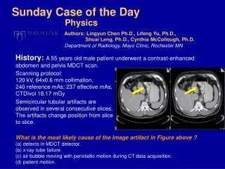
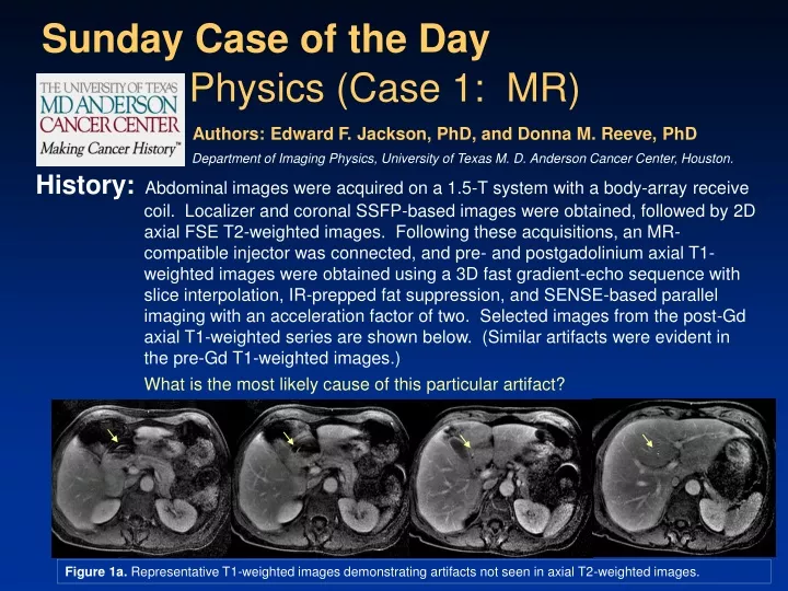
History:Abdominal images were acquired on a 1.5-T system with a body-array receive coil. Localizer and coronal SSFP-based images were obtained, followed by 2D axial FSE T2-weighted images. Following these acquisitions, an MR-compatible injector was connected, and pre- and postgadolinium axial T1-weighted images were obtained using a 3D fast gradient-echo sequence with slice interpolation, IR-prepped fat suppression, and SENSE-based parallel imaging with an acceleration factor of two. Selected images from the post-Gd axial T1-weighted series are shown below. (Similar artifacts were evident in the pre-Gd T1-weighted images.) What is the most likely cause of this particular artifact? Sunday Case of the Day Physics (Case 1: MR) Authors: Edward F. Jackson, PhD, and Donna M. Reeve, PhD Department of Imaging Physics, University of Texas M. D. Anderson Cancer Center, Houston. Figure 1a. Representative T1-weighted images demonstrating artifacts not seen in axial T2-weighted images.
Findings:A more complete set of T1-weighted images (with artifact) and matching T2-weighted images without similar artifact are provided below. Figure 1b. Axial T1-weighted post-Gd images (top) and corresponding axial T2-weighted images (bottom).
Diagnosis:The artifacts are due to aliasing in the SENSE parallel imaging reconstruction. Signal from contrast agent in the coiled MR-compatible injector tubing that was, in this particular case, inappropriately positioned across the patient’s abdomen was aliased into the image.
Aliased Images SENSE: unfold Low-resolution fully encoded coil sensitivity map Unaliased Image 4 3 2 1 Discussion:Parallel imaging techniques accelerate image acquisition rates by utilizing coil sensitivity modulation to encode the MR image and reduce the number of encoding steps. Sensitivity Encoding (SENSE) is one approach to parallel imaging that requires the acquisition of a coil sensitivity reference scan prior to a reduced-phase FOV imaging series. The reduced-phase FOV results in purposeful aliasing of the image in the phase-encode direction. During the reconstruction process, the sensitivity information from the multielement coil is used to “unwrap” the aliased images. Information outside the reconstructed FOV, however, commonly aliases into the FOV, as it cannot be appropriately unwrapped in the reconstruction process. In this particular case, the coiled contrast agent delivery tube, resting on top of the anterior portion of the body array, gave rise to signal that was aliased into the anatomy because it was outside the field of view of the reconstructed image. The T2-weighted images and any images acquired without SENSE parallel imaging reconstruction did not demonstrate such artifacts.
Reference/Bibliography: Pruessmann KP, Weiger M, Scheidegger MB, Boesiger P. SENSE: sensitivity encoding for fast MRI. Magn Reson Med 1999;42:952–962.