Download
1 / 35
350 likes | 382 Views
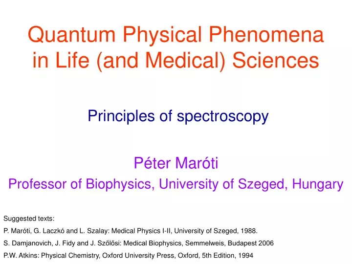
Quantum Physical Phenomena in Life (and Medical) Sciences. Principles of spectroscopy. Péter Maróti Professor of Biophysics, University of Szeged, Hungary. Suggested texts: P. Maróti, G. Laczkó and L. Szalay: Medical Physics I-II, University of Szeged, 1988.

E N D
Quantum Physical Phenomena in Life (and Medical) Sciences Principles of spectroscopy Péter Maróti Professor of Biophysics, University of Szeged, Hungary Suggested texts: P. Maróti, G. Laczkó and L. Szalay: Medical Physics I-II, University of Szeged, 1988. S. Damjanovich, J. Fidy and J. Szőlősi: Medical Biophysics, Semmelweis, Budapest 2006 P.W. Atkins: Physical Chemistry, Oxford University Press, Oxford, 5th Edition, 1994
Definition of the spectrum Spectrum: distribution of a physical quantity according to energy. Physical quantities can be e.g. emitted or absorbed light quanta (photons) Energy can be represented by wavelength, frequency, wave number etc. Example: the absorption spectrum. Selective absorption of the light by visual pigments of the cones and rod in the human eye. This is necessary to the color vision.
Electromagnetic spectrum visible light high energy low energy energy frequency radio microwave infrared ultraviolet X-ray wavelength
Magnetic Resonance SpectroscopyElectron Spin Resonance (ESR)or Electron Paramagnetic Resonance (EPR)andNuclear Magnetic Resonance (NMR)
NMR ~10-200 MHz @ 4.7 T mI= -½ EPR ~9.5GHz @ 0.34 T mI= ±½ Energy (n) RT=0.002 kcal/mol at 10oK Radiowave Energy of transition n =E/h (E = hn) mI= +½ 0 magnetic field strength (B0) ms= -½ RT=0.593 kcal/mol at 298oK ms= ±½ Energy (n) n - frequency Hz n - wavenumber cm-1 • - wavelength nm Microwave ~ ms= +½ 0 magnetic field strength (B0)
EPR and NMR spectroscopy 1) EPR spectrum: absorbed electromagnetic radiation as a function of the magnetic induction. 2) The magnitude of the applied magnetic induction is smaller than in EPR but electromagnetic radiation of larger frequency (microwave) is needed. 3) Spin-marker: compounds with stable and unpaired electrons. 4) Kinetic measurements: to track translation and rotation in the ms time domain. 1) NMR spectrum: absorbed electromagnetic radiation as a function of the magnetic induction. The area under the line (band) of the spectrum is proportional to the number of the absorbing nuclea. 2) The position of the lines depends on the interactions („chemical shift”): the structure of the electron clouds modifies the local magnetic field (induction) and therefore the condition for resonance absorption is shifted. Excellent tool for determination of the chemical structure. The protons in three positions offer three different groups of lines. CH2 OH CH3 quartet singlet triplet Magnetic induction (gauss, 100 μT) Chemical shift
Magnetic Resonance Spectroscopy Electron Paramagnetic Resonance (EPR) spectroscopy • Electrons have “spin”, - rotation of the charge about its axis generates a magnetic field at each electron. • Electrons in orbitals with two electrons are spin-paired, - one with ms+½ (, a), one with ms -½ (, b), - so that the spins and magnetic fields cancel (). • Most molecular bonds are formed by coalescence of atomic orbitals so as to satisfy the lower energy state arising from spin-pairing. Most molecules therefore have all orbitals occupied by magnetically “silent” electron pairs. • If an electron is added to or subtracted from such a molecule (by reduction or oxidation) an unpaired electron is introduced which is not spin coupled, and therefore acts as a magnet. Such molecules are said to be paramagnetic. • Some bonds have unpaired electrons, giving them an inherent paramagnetic property. • In EPR spectroscopy, we take advantage of this paramagnetic property to measure properties of the the paramagnetic center, and its interaction with other local magnets. These interactions provide information about local structure, including local environment, distances, angles, polarity, etc.
Electron Paramagnetic Resonance ms= -½ The photon energy for resonance depends on the applied field. The field aligns the spins, to give a splitting of the electron energy levels, with a population in the ms +½ state that is greater than that in the ms -½ state. Absorption of a photon at the resonance energy flips the spins. The net absorbance is due to transitions from ms +½ to ms -½ levels. ms= ±½ Energy (n) ms= +½ 0 magnetic field strength (B0) Absorption of microwaves changes the energy level so that the small fraction of spins in the lower energy state are flipped into the opposite orientation. Saturation occurs when flips in both directions occur with equal probability. The power needed to saturate depends on the relaxation time of the spin transition. Relaxation No field External field applied
The nomenclature for EPR The general principles of EPR and NMR are essentially the same, but with the differences in energies. However, the nomenclature is different, mainly for historical reasons. The resonance condition for EPR is described by the equations: Here, g, the dimensionless Landég-value, describes the resonance energy. Values around 2.003 are found for simple free-radical systems. The term b(written also in form of μB)is the Bohr magneton: b = eh/(4p me)=927.4 x 10⁻²⁶ JT-1 in SI units, or bn = b/h = e/(4p me) = 13.996 x 10⁹ Hz T-1 in frequency units, or beV = b /(e/C) = 5.7884 x 10⁻⁵ eV T-1 in electron volt units, where (e/C) is the electron charge, 1.602 x 10⁻¹⁹ C.
Nuclear Magnetic Resonance (NMR) spectroscopy • The atomic nucleus is made up of protons (+ charge) and neutrons (neutral). • Like electrons, protons and neutrons (or nucleons) are quantum mechanical entities, and their energetic properties can be described by operators, wave functions, and quantum numbers. • Both protons and neutrons have a spin ½, with spin quantum number, mI, which can have values of (up, +½, or a), or (down, -½, or b). • Total nuclear energy levels are lower if the constituent protons and neutrons are spin-paired, so that for most nuclei, the spins (and magnetic fields) cancel. Protons are spin-paired with protons, neutrons with neutrons. • Isotopes that have an odd number of either protons or neutrons have a net spin, according to the Table below:
Nuclear Magnetic Resonance mI= -½ In the absence of an external magnetic field, the spins of the nuclei are arranged more or less at random. When a magnetic field is applied, the spins align with the field, just like bar magnets. However, because the energy involved is so small, they can flip direction relatively easily. mI= ±½ Energy (n) mI= +½ 0 magnetic field strength (B0) The consequence is that the energy level of the nucleus splits in a magnetic field, as shown on the right. The population of spins in the mI +½ (a) state is (very slightly) higher than in the mI -½ (b) state, because the energy is lower.
Resonance conditions at NMR When photons of the “right” energy are absorbed, the spin of the nucleus flips between the two states. If we look at the diagram, we can see that the energy gap between the two states is dependent on the strength of the applied magnetic field (magnetic induction, B0). As a consequence, photons are absorbed which have a particular energy at a particular field. This is what is meant by resonance. Here, g is the gyromagnetic ratio for the nucleus, B0 is the magnetic induction, and h is the Planck’s constant. When we come to discuss pulsed NMR, we will need to refer to circular motion. Here the preferred frame of reference is that of the circular motion of precession. We define the precession or Larmor frequency:
Some consequences of the energy scale. The transitions leading to NMR absorption have energies in the radio frequency range, depending on nucleus (g) and the strength of the magnetic field generated by the magnet. NMR machines are rated by the frequency at which the proton is in NMR resonance for the magnet they are built around, so we have 200 MHz, 500 MHz, 750 MHz and even 1 GHz NMR spectrometers. To achieve the higher fields, high electrical currents are needed, which can be achieved using superconducting coils, - these are generally called superconducting magnets (costing $M). Since w = 2pn = gB0, for a 200 MHz machine we need a magnet generating 4.7 Tesla; for a 500 MHz machine we need 11.74 Tesla, etc. As we increase the energy gap (increase frequency), the small differences of energy for transitions of nuclear magnets in different environments are also increased, and our NMR spectrum will be better resolved. In addition, we also increase the population difference for the two spin states as we increase the energy (DE/RT is increased), as discussed in the next slide. These factors make a big difference in the amount of time needed to generate a data set, - for example in solution of the structure of a protein.
As we have seen, the energy of electromagnetic waves is generally expressed in frequency, but energy scales are all related through Planck’s constant and the speed of light, so we can express these energies in J/mol, eV, or any other energy units. For a particular temperature, any transition can be described in terms of an equilibrium between two states. For example, when we flip the energy level of a proton in a magnetic field, we have two states, or a, and or b, separated by DE, calculated as above. Let us use Na and Nb to represent the relative populations in the two states. We can represent the equilibration between these two states by NaNb . Then the ratio of these populations is given by the Boltzman distribution: Na / Nb = e DE / RT Remembering that the energies of the transitions associated with NMR (and those of EPR) are both very much less than RT, we can see that only a very small excess of states will be in the lower energy level. For the proton at 500 MHz, at ~25oC, since E = hn, and Planck and Avagadro constants are 6.626.10-34 J.s and 6.02.1023 mol-1 respectively, energies in the RF range (~500 MHz) are 0.2 J/mol. Na / Nb= 1.00008 It is these one in ten thousand spins that are available to absorb a photon to provide an NMR signal.
Molecular vibrationsVibrational spectroscopy Frederick William Herschel (~1800) He discovered the non-visible (infrared) part of the spectrum of the sun.
Elektromagnetic spectrum X-RAY 0.2 nm ULTRA-VIOLET 2 nm VISIBLE 400-800 nm MICROWAVE 3 mm-20 cm RADIO 10 m-30 km INFRARED NEAR IR Mid IR FAR IR wavelength (cm)7.8·10-5 to 3·10-4 3·10-4 to 3·10-3 3·10-3 to 3·10-2 wavenumber (cm-1)12820 to 4000 4000 to 400 400 to 33 Information about the molecules Overtones and combinations of the normal vibrations Basic (normal) vibrations Rotations
3. Particles (atoms and molecules) in parabolic potential well The energy profile of a harmonic oscillator is parabolic. The particle is freely moving inside the parabolic potential well. Semiclassic treatment The total energy of the particle is which is equal to the kinetic energy between the walls. Those energy levels are allowed which can be covered with standing waves Possible velocities of the particle Possible energies of the particle
Energy levels of the harmonic oscillator Uniform ladder of spacing h·ν Solution from the Schrödinger equation: En = h·ν(n + ½), where n = 0, 1, 2, ... • Conclusions: • The energy levels of the oscillator are equidistant: ΔE = h·ν. • The selection rule: Δn = ± 1. • Transitions are allowed between adjacent states only (in first order). Transitions among not neigboring levels are forbidden. • (c) The minimum energy of the oscillator (n = 0) differs from zero: E0 = ½ hν. The zero-point energy is not zero, because of the uncertainty principle: the particle is confined, its position is not completely uncertain, and therefore its momentum, and hence its kinetic energy, cannot be exactly zero. For typical molecular oscillator, the zero-point energy is about E0 = 3·10-20 J = 100 meV The Boltzmann-energy at room temperature is ½ kBT = 25 meV
Infrared (IR) spectroscopy • The energy levels are associated with vibrations of bonds in molecules. • Each bond type will absorb IR light at one wavelength only, but there are different modes of vibrations: • valence vibrations and • straching vibrations. All (arbitrarily complex) vibrations of a molecule can be decomposed into the sum of normal modes. Example: normal modes of vibrations of CO2
Normal vibrations antiszimmetric szimmetric R R H H stretch R H R H R R H H in plane bending R H R H rocking scissoring R R H H R H R H out of plane bending
Measuring methods IR Reflexion IR Transmission Detector IR Reflexion IR Transmission IR light IR Absorption IR light Detector
Advantages of the IR spectroscopy Non-invasive determination of the sucrose content of the blood
Advantages of the IR spectroscopy Remote sensing and measurement of hazardous materials IR light source Grating Sample Detector
Fourier transform (FT) techniques in the infrared (IR) spectrometers (FTIR) The heart of the FTIR spectrometer is a Michelson interferometer which works by splitting the beam from the sample into two and introducing a varying path difference x into one of the two arms. When the two components recombine, there is a phase difference between them, and they interfere either constructively or destructively depending on the extra path that one has taken. The detected signal oscillates as the two components alternately come into and out of phase as the path difference is changed. If the radiation has wavenumber k (=1/λ), the detected signal varies with x as The interferometer converts the presence of a particular component in the signal into a variation in intensity of the radiation reaching the detector. An actual signal consists of radiation spanning a large number of wavenumbers, and the total intensity at the detector is the sum of all their oscillating intensities The problem is to find I(k), the variation of intensity with wavenumber, which is the spectrum we require, from the record of values I(x). The step is a standard technique of mathematics: Fourier transformation. known unknown
Raman spectroscopy • About 1 in 107 of the incident laser photons collide with the molecules in the sample, give up some of their energy to the molecular vibrations, and emerge with a lower energy (Stokes radiation). • Other incident photons may collect energy from the molecular vibrations, and emerge as higher-frequency (anti-Stokes) radiation. • Basic properties: • The shifts in frequency are small (good monochromator is needed) and • - the intensities of the lines are low (high laser energy is needed). An advantage of Raman spectroscopy over infrared spectroscopy is that the radiation can be entirely in the visible range, so the complications arising from needing to select a range of infrared-transparent sample cells are avoided.
Comparison of Raman and IR spectroscopies:advantages and disadvantages The two methods utilize complementer properties of the same molecule.
Complexity and complementarity of the infrared (IR) and the Raman spectra Hoop: out-of-plane motion of H atoms streching of the methyl group wavenumber, 1/cm rocking of the methyl group Single bond vibrations deformation of the methyl group Double bond vibrations
Electron Transitionsto rebrush your mindsee the previous chapters on „Electron in hyperbolic and rectangular shape of potential wells” andfor additional informationsee the next slide-show on „Visible absorption and fluorescence spectroscopy: Luminescence”
Problems for Seminar • An electron is confined to a molecule of length 1.0 nm. What is (a) its minimum energy and (b) the minimum excitation energy from that state? • Estimate a typical nuclear excitation energy by calculating the first excitation energy of a proton (mass of m = 1.673·10-27 kg, the Planck’s constant is h = 6.626·10-34 J·s) confined to a region roughly equal to the diameter of a nucleus, 1·10-14 m. • Calculate the zero-point energy of a harmonic oscillator consisting of a particle of mass 2.33·10-26 kg and force constant 155 N/m. • For a harmonic oscillator consisting of a particle of mass 1.33·10-25 kg, the difference in adjacent energy levels is 4.82·10-21 J. Calculate the force constant of the oscillator. • Calculate the minimum excitation energies of (a) a pendulum of length 1.0 m on the surface of the Earth, (b) the 33 Hz quartz crystal of a watch and (c) the bond between two oxygen atoms in the O2 molecule, for which the force constant is k = 1177 N/m.
Problems for Seminar 6. Determine the wavefunction and the probability distribution of the harmonic oscillator in the first excited state. Compare these functions with those of the ground state. 7. Calculate the longest and the shortest wavelength transitions in the Balmer series of atomic hydrogen. 8. Do the same with the Lyman and Paschen series of the H atom. 9. Calculate the saparation between the two lowest levels for an oxygen molecule in a one-dimensional container of length 5.0 cm. At what value of n does the energy of the molecule reach 1/2·kBT at 300 K, and what is the separation of this level from the one immediately below? 10. To a crude first approximation, a π-electron in a linear polyene may be considered to be a particle in a one-dimensional box. The polyene β-carotene contains 22 conjugated C atoms, and the average internuclear distance is 140 pm. Each state up to n = 11 is occupied by two electrons. Calculate the separation in energy between the ground state and the first excited state in which one electron occupies the state with n = 12. What is the wavelength of the radiation required to produce a transition between these two states?
Problems for Seminar 11. The inverse value of the reduced (effective) mass of a diatomic molecule is the sum of the inverse values of the individual masses of the atoms mA and mB: 1/μ= 1/mA + 1/mB. The table contains the infrared absorption wavenumbers (in cm-1) of the following diatomic molecules: Calculate the force constants of the bonds and arrange them in order of increasing stiffness. 12. An Ar atom rotates in a circle around a fixed axis (z) with orbital angular momentum quantum number ml = 2. Its energy of rotation is Erot = 2.47·10-23 J. Calculate the radius (r) of the rotation. (The angular momentum about the axis (Jz = r·p) is quantized: Jz = ml·(h/2π).)