Download
1 / 49
540 likes | 731 Views
Lectures on Medical Biophysics Dept. Biophysics, Medical faculty, Masaryk University in Brno. Biological membranes and bioelectric phenomena.

E N D
Lectures on Medical BiophysicsDept. Biophysics, Medical faculty, Masaryk University in Brno Biological membranes and bioelectric phenomena A part of this lecture was prepared on the basis of a presentation kindly provided by Prof. Katarína Kozlíková from the Dept. of Medical Biophysics, Medical Faculty, Comenius University in Bratislava
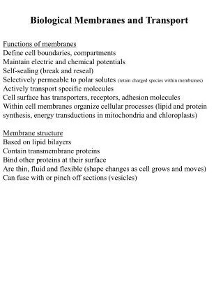
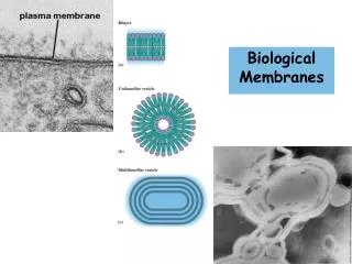
Biological membrane • It is not possible to understand the origin of resting and action membrane voltage (potential) without knowledge of structure and properties of biological membrane. • In principle, it is an electrically non-conducting thin bilayer (6-8 nm) of phospholipid molecules. There are also built-in macromolecules of proteins with various functions. Considering electrical phenomena, two kinds of proteins are the most important: the ion channels and pumps. In both cases these are components of transport mechanisms allowing transport of ions through the non-conducting phospholipid membrane.
Bioelectric phenomena • The electric signal play a key role in controlling of all vitally important organs. They ensure fast transmission of information in the organism. They propagate through nerve fibres and muscle cells where they trigger a chain of events resulting in muscle contraction. They take a part in basic function mechanisms of sensory and other body organs. • On cellular level, they originate in membrane systems, and their propagation is accompanied by production of electromagnetic field in the ambient medium. • Recording of electrical or magnetic signals from the body surface is fundamental in many important clinical diagnostic methods.
Structure of the membrane Phospholipid bilayer Integral proteins
Channels • The basic mechanism of the ion exchange between internal and external medium of the cell are the membrane channels. They are protein molecules but, contrary to the pumps with stable binding sites for the transmitted ions, they form water-permeable pores in the membrane. Opening and closing of the channels (gating) is performed in several ways. Besides the electrical gating we can encounter gating controlled by other stimuli in some channels (chemical binding of substances, mechanical tension etc.). • The passage of ions through the channel cannot be considered to be free diffusion because most channels are characterised by certain selectivity in ion permeability. Sodium, potassium, calcium or chloride channels are distinguished. • In this kind of ion transport there is no need of energy delivery.
Electrical and chemical gating polarised membrane depolarised closed channel open channel closed open channel channel
Ion transport systems • Many ion transport systems were discovered in cell membranes. One of them, denoted as sodium-potassium pump (Na/K pumpor Na+-K+-ATP-ase) has an extraordinary importance for production of membrane voltage. It removes Na-ions from the cell and interchanges them with K-ions. Thus, the concentrations of these ions in the intracellular and extracellular medium (they are denoted as [Na+], [K+] and distinguished by indexes i, e) are different. We can write: Working Na/K pump requires constant energy supply. This energy is delivered to the transport molecules by the adenosine triphosphate (ATP) which is present in the intracellular medium.
Principle of the sodium-potassium pump The sodium ions are released on the outer side of the membrane. Following conformation change of the ion pump molecule enables binding of potassium ions which are carried inside the cell.
Function of biological membranes • They form the interface between the cells and also between cell compartments. • They keep constant chemical composition inside bounded areas by selective transport mechanisms. • They are medium for fast biochemical turnover done by enzyme systems. • Their specific structure and selective ion permeability is a basis of bioelectric phenomena.
Excitability Characteristic feature of living systems on any level of organisation of living matter An important condition of adaptation of living organisms to environment An extraordinary ability of some specialised cells (or tissues – muscle cells, nerve cells) Each kind of excitable tissue responses most easily on a certain energetic impulse (the adequate stimulus). Another energetic impulse can also evoke an excitation but much more energy is necessary (the inadequate stimulus).
Resting membrane potential – RMP (1) Potential difference between a microelectrode inside the cell (negative potential) and a surface electrode outside the cell (zero potential)= membrane voltage = membrane potential „Non-polarisable“ electrodes are used Extracellular space membrane Intracellular space membrane Extracellular space
Resting membrane potential – RMP (2) Its values depend on: - Type of the cell - Art of the animal the cell is taken from - For identical cells – on the composition and concentration of the ion components of the extracellular liquids The value of RMP at normal ion composition of the IC and EC liquid:-100 mV to -50 mV Membrane thickness ~ 10 nm Electric field intensity in the membrane ~ 107 V/m Electric field intensity on the Earth’s surface ~ 102 V/m
Approach to the RMP • (1): Electrodiffusion models: They describe processes phenomenologically on the basis of thermodynamics. Origin of the RMP is connected with diffusion of ions across the membrane - Nernst and Donnan models, ion transport model • (2): Physical – based on description of behaviour of solids or liquid crystals • describe movement of ions across the membrane and its blocking • they consider characteristic propertiesof structural elements of the membrane (lipids, proteins) • (3) Models based on equivalent electrical circuits: They describe behaviour of the cells in rest or excited state. Electrical properties of the cells are considered in accord with other models.
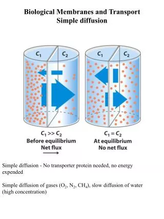
Diffusion potential DP (1) Caused by diffusion of charged particlesDP in non-living systems – solutions are separated by a membrane permeable for Na+ and Cl-. Electric field repulses Cl- from [2] Hydration envelope (water molecules are bound to ions) Na+(more) a Cl-(less) faster diffusion of Cl- against (!) concentration gradient Transient voltage appears across the two compartments Diffusion potential The compartments are electroneutral, but there is a concentration gradient Diffusion of ions from [1] do [2]
Diffusion potential DP (2) DP in living systems – the solutions are separated by a selectively permeable membrane for K+, non-permeable for pro Na+ a Cl-. Diffusion of K+ against its concentration gradient occurs until an electric gradient of the same magnitude, but of opposite directionarises An equilibrium potential emerges – resulting diffusion flux is equal to zero In such a system, an equilibrium arises if there is no resulting flux of ions.
A simple example of a membrane equilibrium (1) The same electrolyte is on both sides of the membrane but of different concentrations (cI > cII), the membrane is permeable only for cations membrane Result: Electric double layer is formed on the membrane layer 1: anions stopped in space I layer 2: cations attracted to the anions (II) Electrolyte II Electrolyte I cations cCI anions cAII anions cAI cations cCII
A simple example of a membrane equilibrium (2) The concentration difference ”drives” the cations, electric field of the bilayer “pushes them back” In equilibrium: potential difference U arises: membrane Electrolyte II Electrolyte I - - - - - - - - - + + + + + + + + + I II cations cCI anions cAII anions cAI cations cCII (Nernst equation)
Donnan equilibrium (1) The same electrolyte is on both sides, concentrations are different (cI > cII), membrane is permeable for small univalent ions C+ and A-, non-permeable for R-. membrane Diffusible ions:C+, A- diffuse freely non-diffusible ions:R- Electrolyte I Electrolyte II anions R- In presence of R-: Equal distribution of C+ and A- cannot be achieved a special case of equilibrium - Donnan equilibrium cations cCI anions cAII anions cAI cations cCII
Donnan equilibrium (2) Equilibrium concentrations: membrane Donnan ratio: Electrolyte I Electrolyte II anions R- cations cCI anions cAII anions cAI cations cCII
Donnan equilibrium (3) Donnan ratio: membrane Donnan potential: Electrolyte I Electrolyte II + + + + + + + + + + + - - - - - - - - - - - anions R- cations cCI anions cAII anions cAI cations cCII
Donnan model in living cell (1) diffuse: K+, Cl- do not diffuse: Na+, anions, also proteins and nucleic acids cell membrane intra extra Concentrations: [K+] in > [K+] ex [Cl-] in < [Cl-] ex phosphate anions Na+ protein anions K+ Cl- Cl- K+
Donnan model in living cell (2) Donnan ratio: Cell membrane Donnan potential: intra extra - - - - - - - - - - - + + + + + + + + + + + phosphate anions Na+ protein anions K+ Cl- Cl- K+
Donnan model in living cell (3) Donnan potential (resting potential) [mV]: object: calculated: measured: K+: Cl-: cuttlefish axon - 91 - 103 - 62 frog muscle - 56 - 59 - 92 rat muscle - 95 - 86 - 92 • Donnan model differs from reality: • The cell and its surroundings are regarded as closed thermodynamic systems • The diffusible ions are regarded as fully diffusible, the membrane is no barrier for the diffusible ions • The effect of ionic pumps on the concentration of ions is neglected • The interaction between membrane and ions is not considered
Model of ion transport (1) Electrodiffusion model with smaller number of simplifications. We suppose: A constant concentration difference between outer and inner side of the membrane constant transport rate through membrane Migration of ions through membrane electric bilayer on both sides of the membrane All kinds of ions on the both sides of the membrane are considered simultaneously Empirical fact – membrane is neither fully permeable nor fully non-permeable for any ion Different ions have different permeability
Model of ion transport (2) Goldman - Hodgkin - Katz k = cations, a = anions P - permeability
Model of ion transport (3) „giant“ cuttlefish axon (t = 25°C): pK : pNa : pCl = 1 : 0.04 : 0.45 calculated: U = - 61 mV measured: U = - 62 mV frog muscle (t = 25°C): pK : pNa : pCl = 1 : 0.01 : 2 calculated: U = - 90 mV measured: U = - 92 mV
Action potential
Action potential • The concept of action potential denotes a fast change of the resting membrane potential caused by over-threshold stimulus which propagates into the adjacent areas of the membrane. • This potential change is connected with abrupt changes in sodium and potassium ion channels permeability. • The action potential can be evoked by electrical or chemical stimuli which cause local decrease of the resting membrane potential.
Mechanism of action potential triggering Um t UNa AP 0 Depolarization phase Positive feedback: gNa depol gNa Repolarization phase: inactivation gNa and activation gK Upr Umr t UK hyperpolarization (deactivation gK) Mechanism of the action potential triggering in the cell membrane is an analogy of a monostable flip-flop electronic circuit.
Action potential • Changes in the distribution of ions caused by action potential are balanced with activity of ion pumps (active transport). • The action potential belongs among phenomena denoted as „all or nothing“ response. Such response is always of the same size. Increasing intensity of the over-threshold stimulus thus manifests itself not as increased intensity of the action potential but as an increase in action potential frequency (rate).
Propagation of the action potential along the membrane AP propagation is unidirectional because the opposite side of the membrane is in the refractory period.
Propagation of AP and local currents time AP propagates along the membrane as a wave of negativity by means of local currents
Conduction of action potential along the myelinated nerve fibre Saltatory conduction
Examples of action potentials A–nerve fibre, B – muscle cell of heart ventricle; C – cell of sinoatrial node; D – smooth cell muscle.
Definition • Synapse is a specific connection between two neurons or between neurons an other target cells (e.g. muscle cells), which makes possible transfer of action potentials. We distinguish: • Electrical synapses (gap junctions) – close connections of two cells by means of ion channels. They enable a fast two-way transfer of action potentials. • Chemical synapses – more frequent, specific structures, they enable one-way transfer of action potentials.
Transmission of action potential between neurons
Chemical synapse – electron micrograph Mitochondrion Vesicles Synaptic gap (cleft)
Synaptic mediators (neurotransmitters) • The most frequent mediators (neurotransmitters) of excitation synapses areacetylcholine(in neuromuscular end plates and CNS) andglutamic acid(in CNS). Both compounds act as gating ligands mainly for sodium channels. Influx of sodium ions inside the cell evokes a membrane potential change in positive sense – towards a depolarisation of the membrane (excitation postsynaptic potential). • Gamma-amino butyric acid (GABA) is a neurotransmitter of inhibitory synapses in brain. It acts as a gating ligand of chloride channels. Chloride ions enter the cell and evoke so a membrane potential change in negative sense – membrane hyperpolarization results (inhibitory postsynaptic potential)
Excitation and inhibition postsynaptic potential
Summation of postsynaptic potentials
Summary • Electric phenomena on biological membranes play a key role in functioning of excitatory tissues (nerves, muscles) • Resting membrane potential (correctly: membrane voltage) is a result of a non-equal distribution of ions on both sides of the membrane. • It is maintained by two basic mechanisms: selective permeable ion channels and by transport systems – both these systems have protein character • Changes of membrane voltage after excitation are denoted as action potentials • Membrane undergoes two phases after excitation: depolarization – connected with influx of sodium iions into the cell - and subsequent repolarization – connected with efflux of potassium ions from the cell • In the refractory period, the membrane is either fully or partly insensitive to stimulation • Synapse is a connection of two cells which enables transmission of action potentials
„Only two things are infinite, the universe and human stupidity, and I am not sure about the former“. Albert Einstein (1879-1955)
Authors: Vojtěch Mornstein, Ivo HrazdiraLanguage collaboration: Carmel J. CaruanaPresentation design: - Last revision: February 2012