Download

1 / 19
190 likes | 194 Views
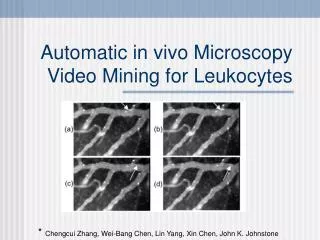
This paper proposes an automatic video mining approach to track and calculate the velocity of moving leukocytes and detect the magnitude of adherent leukocytes in in-vivo microscopy images. It addresses challenges such as server noise, background movement, and contrast changes. Two approaches, probabilistic learning and neural network, are used for detecting moving leukocytes, and a polynomial fitting method is employed for detecting adherent leukocytes. Experimental results show promising accuracy.

E N D
Automatic in vivo Microscopy Video Mining for Leukocytes * Chengcui Zhang, Wei-Bang Chen, Lin Yang, Xin Chen, John K. Johnstone
Background Information • What is in vivo microscopy? • Images of the cellular and molecular processes in a living organism • Why video-mine leukocytes? • To Predict Inflammatory response • Rolling velocity and magnitude of adhesion of leukocytes are the main predictors • Currently analyzed manually • Time consuming / Expensive • Subjective
Objectives • Given a sequence of in vivo images, • Track the moving leukocytes • Calculate their average velocity • Find the magnitude of adherent leukocytes
Challenges • Server Noise • Background movement • Due to movement of the living organism • Deformation of leukocytes • Change of contrast in different frames
Previous Work • [Eden et al.] use local features (e.g. color) for a tracking system • Assume that leukocytes roll along the vessel centerline • [Acton et al.] Background removal + morphological filter • Assumes the shape/size leukocytes does not change
Suggested Approach • Three main steps: • Frame Alignment • To correct the camera/subject movement • Detect Moving Leukocytes • Detect Adherent Leukocytes • After moving leukocytes are removed
Step 1- Frame Alignment • 1.1- Detect Camera/Subject Movement • Define a (dis)similarity measure between consecutive frames • This allows for some tolerance within radius r • If S(ft-1, ft) is larger than a threshold, then ft requires frame alignment
Step 1- Frame Alignment • 1.2- Frame Matching • Generate a number of high dimensional, local scale-invariant features [SIFT] for the frame and its predecessor • Use nearest-neighbor to find a match for each feature point • Calculate the transformation matrix H, such that • For every matched point x and x’
Step 1- Frame Alignment • Use Random Sample Consensus (RANSAC) to correct the mismatches
Step 2 - Detecting Moving Leukocytes • Approach 1 - Probabilistic Learning • For pixel j in the image, let x1j, x2j, ..., xNj be the intensity of the pixel over N frames • Assume that P(xtj) has a normal distribution over time with mean xtj • If P(xtj) is smaller than a threshold, then it is a foreground pixel • Problem: Difficult to find a threshold
Step 2 - Detecting Moving Leukocytes • Approach 1 - Probabilistic Learning • Problem: Difficult to find the threshold value • Solution: Use One-Class SVM to classify background and foreground pixels
Step 2 - Detecting Moving Leukocytes • Approach 2 - Neural Network • Train a neural net to learn the predictable pattern of the background pixels • Input: [x(t-m), x(t-m+1),... , x(t-1)] • A sliding window of the intensity sequence • Output: x(t) • Prediction for the intensity of the pixel at the next frame • If the neural-net prediction and the real pixel intensity are very different, the pixel in the current frame is in foreground
Step 2 - Detecting Moving Leukocytes • Approach 2 - Neural Network
Step 2 - Detecting Moving Leukocytes • Calculating the leukocytes velocity • Find the centroid of each group of connected foreground pixels • For each centroid, find the closest centroid in the previous frame • If their distance is smaller than a threshold, they are a match • Compute the mean velocity
Step 3- Detecting Adherent Leukocytes • First, remove the moving leukocytes • Three main types of regions left • Tissues • Vessels • Adherent Leukocytes • These three have different intensity values
Step 3- Detecting Adherent Leukocytes • Finding the threshold values • Fit an 8th degree polynomial to the histogram curve • The real part of the second largest root is the ideal threshold • Justification? • Problem with false positives and false negatives
Experimental Results • Test video of 148 frames • Detecting moving leukocytes: • 1% false positive for probabilistic learning(?) • 49% false positive for neural-net approach • 50% recall • Detecting Adherent leukocytes • 2% false positive • 95% recall
Final Remarks • Paper is mainly related to Vision • The algorithms require many “magic parameters” that need hand tuning • Would the current parameters work as well for a new video sequence from a new equipment? • Do we want to pursue more video-mining papers?