Download
1 / 16
440 likes | 1.38k Views
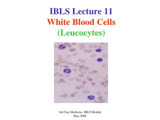
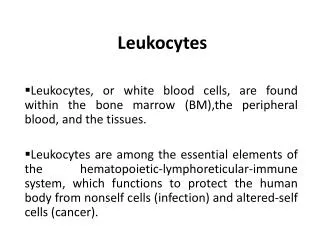
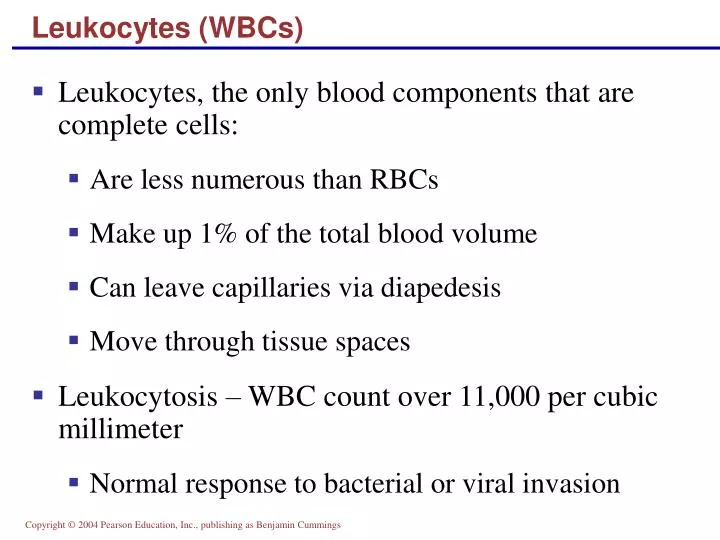
Leukocytes (WBCs). Leukocytes, the only blood components that are complete cells: Are less numerous than RBCs Make up 1% of the total blood volume Can leave capillaries via diapedesis Move through tissue spaces Leukocytosis – WBC count over 11,000 per cubic millimeter

E N D
Leukocytes (WBCs) • Leukocytes, the only blood components that are complete cells: • Are less numerous than RBCs • Make up 1% of the total blood volume • Can leave capillaries via diapedesis • Move through tissue spaces • Leukocytosis – WBC count over 11,000 per cubic millimeter • Normal response to bacterial or viral invasion
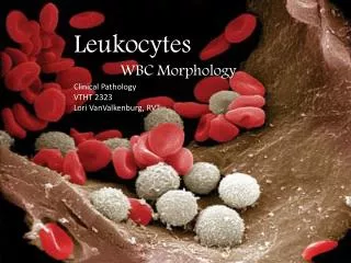
Granulocytes • Granulocytes – neutrophils, eosinophils, and basophils • Contain cytoplasmic granules that stain specifically (acidic, basic, or both) with Wright’s stain • Are larger and usually shorter-lived than RBCs • Have lobed nuclei • Are all phagocytic cells
Neutrophils • Neutrophils have two types of granules that: • Take up both acidic and basic dyes • Give the cytoplasm a lilac color • Contain peroxidases, hydrolytic enzymes, and defensins (antibiotic-like proteins) • Neutrophils are our body’s bacteria slayers
Eosinophils • Eosinophils account for 1–4% of WBCs • Have red-staining, bilobed nuclei connected via a broad band of nuclear material • Have red to crimson (acidophilic) large, coarse, lysosome-like granules • Lead the body’s counterattack against parasitic worms • Lessen the severity of allergies by phagocytizing immune complexes
Basophils • Account for 0.5% of WBCs and: • Have U- or S-shaped nuclei with two or three conspicuous constrictions • Are functionally similar to mast cells • Have large, purplish-black (basophilic) granules that contain histamine • Histamine – inflammatory chemical that acts as a vasodilator and attracts other WBCs (antihistamines counter this effect)
Agranulocytes • Agranulocytes – lymphocytes and monocytes: • Lack visible cytoplasmic granules • Are similar structurally, but are functionally distinct and unrelated cell types • Have spherical (lymphocytes) or kidney-shaped (monocytes) nuclei
Lymphocytes • Account for 25% or more of WBCs and: • Have large, dark-purple, circular nuclei with a thin rim of blue cytoplasm • Are found mostly enmeshed in lymphoid tissue (some circulate in the blood) • There are two types of lymphocytes: T cells and B cells • T cells function in the immune response • B cells give rise to plasma cells, which produce antibodies
Monocytes • Monocytes account for 4–8% of leukocytes • They are the largest leukocytes • They have abundant pale-blue cytoplasms • They have purple-staining, U- or kidney-shaped nuclei • They leave the circulation, enter tissue, and differentiate into macrophages
Monocytes • Macrophages: • Are highly mobile and actively phagocytic • Activate lymphocytes to mount an immune response
Summary of Formed Elements Table 17.2
Summary of Formed Elements Table 17.2
Production of Leukocytes • Leukopoiesis is hormonally stimulated by two families of cytokines (hematopoietic factors) – interleukins and colony-stimulating factors (CSFs) • Interleukins are numbered (e.g., IL-1, IL-2), whereas CSFs are named for the WBCs they stimulate (e.g., granulocyte-CSF stimulates granulocytes) • Macrophages and T cells are the most important sources of cytokines • Many hematopoietic hormones are used clinically to stimulate bone marrow
Formation of Leukocytes • All leukocytes originate from hemocytoblasts • Hemocytoblasts differentiate into myeloid stem cells and lymphoid stem cells • Myeloid stem cells become myeloblasts or monoblasts • Lymphoid stem cells become lymphoblasts • Myeloblasts develop into eosinophils, neutrophils, and basophils • Monoblasts develop into monocytes • Lymphoblasts develop into lymphocytes
Formation of Leukocytes Figure 17.11
Leukocytes Disorders: Leukemias • Leukemia refers to cancerous conditions involving white blood cells • Leukemias are named according to the abnormal white blood cells involved • Myelocytic leukemia – involves myeloblasts • Lymphocytic leukemia – involves lymphocytes • Acute leukemia involves blast-type cells and primarily affects children • Chronic leukemia is more prevalent in older people
Leukemia • Immature white blood cells are found in the bloodstream in all leukemias • Bone marrow becomes totally occupied with cancerous leukocytes • The white blood cells produced, though numerous, are not functional • Death is caused by internal hemorrhage and overwhelming infections • Treatments include irradiation, antileukemic drugs, and bone marrow transplants