Download
1 / 22
220 likes | 427 Views
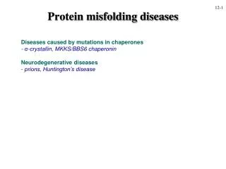
Multiphoton ANS Fluorescence Microscopy as an in vivo Sensor for Protein Misfolding Stress. Tingwei Guo 11-30-2011. Introduction. Cellular stress induced by protein misfolding is at the center of many human diseases. Such as Parkinson’s disease and Huntington’s disease.

E N D
Multiphoton ANS Fluorescence Microscopy as an in vivoSensor for Protein Misfolding Stress Tingwei Guo 11-30-2011
Introduction • Cellular stress induced by protein misfolding is at the center of many human diseases. Such as Parkinson’s disease and Huntington’s disease. • More broadly, elevated burdens of unfolded intracellular protein are linked to malignant transformation and may lie at the foundation of human aging. • Existing methods to measure protein misfolding stress rely on proxy biomarkers for protein folding, chaperone and proteasome function • But these measures generally require the introduction and expression of transgenes or are only suitable for endpoint analyses.
In this paper they have found a novel application of a nontoxic small molecule which binds to intracellular targets and exhibits elevated fluorescence in response to protein-damaging cell stresses. • They employ multiphoton imaging of 1-anilino-8-naphthalenesulfonate (ANS) to noninvasively and quantitatively detect and signal changes in intracellular protein folding. • Fluorescent fusion proteins combining proteins of interest with green fluorescent protein (GFP) or related fluorophores can be expressed to directly track specific, individual disease-associated proteins. • Misfolding of a particular protein can be linked to levels of expression, cellular or tissue localization, or to a portion of a cell or organism’s life cycle.
However, the detection of endogenous proteins or nucleic acids generally requires solubilization or fixation of the cell, preventing the tracking of these markers over time in individual live cells. • They believe that ANS imaged by multiphoton fluorescence microscopy is a probe that is broadly sensitive to the stresses of protein misfolding in vivo. • ANS is relatively nontoxic and can enter all compartments of living cells. Therfore, it avoids transfection or direct modifications of the cell’s biology. • This small molecule is essentially non-fluorescent in aqueous solution, but its fluorescence quantum yield increases several hundredfold in a hydrophobic environment. • In practice, this means that ANS becomes highly fluorescent when embedded in exposed hydrophobic pockets of proteins in partly folded or “molten globule” states.
Historically, ANS has been unsuited to live-cell imaging applications by epifluorescence microscopy, because it is an ultraviolet-excited dye. • It binds strongly to serum albumin, generating an overwhelming background fluorescence under normal mammalian cell culture conditions. • The introduction of multiphoton microscopy opens the door to imaging intracellular ANS fluorescence. • In this paper, they use this technique to image ANS binding sites in living cells.
Results The fluorescence emission spectra of equal mass concentrations of Aβ42 or lipid vesicles in solution with ANS are shown in Fig. 1a. Curve A: Aβ42 at pH 6.0 misfolds into amyloid fibrils and shows a much greater degree of ANS binding and fluorescence. Curve C: At pH 8.0, Aβ exists as a stable, unstructured, soluble species, and shows low fluorescence in the presence of ANS. Curve B: lipid Bilayers prepared from polar lipids. Curve D: synthetic phopholipid. The fluorescence of ANS bound to misfolded Aβ42 is significantly higher than ANS bound to unstructured soluble Aβ42 or lipid bilayers prepared from polar lipids extracted from HeLa cells or from synthetic phospholipid
Results • They wondered whether this vivid response might also occur in the more complex environment of mammalian tissue. • Figure 1b shows a section of transgenic CRND8 mouse brain stained with ANS and imaged by two-photon microscopy. Another section from the same brain is shown stained with thioflavin S (ThS, Fig. 1c), a widely used fluorescent probe specific for amyloid deposits
Results • (Fig. 1d, e), the amyloid plaques are vividly fluorescent. • extended patches of cerebrovascular amyloid lining blood vessels (enlarged in Fig. 1f, g) are also visible. • .Figure 1h shows sections of a low-grade (WHO grade I) pilocytic astrocytoma stained with hematoxylin (Fig. 1h) and ANS (Fig. 1i). • ANS also appears to be sensitive to non-amyloid deposits of aggregated protein that are not detected by thioflavin S
Result • While ANS fluorescence in the tissue sections was the most intense within the amyloid plaques and nonamyloid Rosenthal fibers. • they observed some fluorescence from the surrounding tissue that exhibited normal morphology. • Because there was no conspicuous pathology associated with this other tissue, this finding prompted them to investigate what non-pathological intracellular structures might also bind ANS.
Results • Living HeLa cell stained with ANS under normal growth conditions. • The cell nucleus is clear of ANS fluorescence. • Vivid fluorescence is observed adjacent to the nucleus while regions of both diffuse and punctate ANS fluorescence are seen throughout the cytoplasm.
Results • ANS is not toxic to living cells at the concentrations used for imaging.
Result • We sought to identify the major sites of intracellular ANS binding in unstressed HeLa cells using a variety of fluorescent organelle probes (Fig. 3).
Result • Strong colocalization was observed throughout the cell between ANS fluorescence and a probe for the endoplasmic reticulum (Fig. 3a, f). Bright ANS fluorescence was also observed in the Golgi (Fig. 3b, g). • These organelles are associated with the translation, folding, and/or packaging of cellular proteins.
Results • Fluorescence of the lysosomal probe LysoTracker Green colocalized with brightly ANS-fluorescent cytoplasmic punctae (Fig. 3c, h). • Lysosomes have a low pH, promoting non-native protein conformational states, and are a site of intracellular protein degradation.
Results • In contrast, fluorescence of ANS did not colocalize with a mitochondrial probe (Fig. 3d, i) • nor with intracellular neutral lipid droplets (Fig. 3e, j).
Results • The results show that strong overlap of organelle markers with ANS fluorescence is observed for the endoplasmic reticulum, Golgi, and lysosomes. Poor overlap is noted for the mitochondria, the nucleus, and intracellular neutral lipids • Results indicate that ANS fluorescence is predominantly associated with organelles expected to contain proteins in non-native conformations
Results • Hsp70 is known to reversibly bind to short stretches of hydrophobic residues exposed during de novo protein synthesis or made accessible by stress induced misfolding. • Under normal growth conditions, Hsp70-YFP is predominantly cytoplasmic, but still detectable within the nucleus (Fig. 4d).
Results • Fig. 4 HeLa cells are shown before (a, d) or after (b, c, e, f) linear thermal lesions. • ANS (b, c) bound immediately to the lesion site. • YFP conjugated Hsp70 was also rapidly recruited to the lesion (e), but YFP expressed on its own (f) was not.
Results • Figure 4 shows that authors are generating a misfolded protein that specifically binds the Hsp70 chaperone. • Detecting this damaged protein can use ANS.
Results—heat shock experiment • Nonlethal heat shock drives a dramatic translocation of Hsp70. • Its nucleocytoplasmic distribution is reversed, and the nuclear concentration of Hsp70—particularly near the nucleolus—becomes quite high after heat shock. • Following hyperthermia exposure, nuclei (especially nucleoli) exhibit elevated ANS fluorescence.
Results---proteasome experiment • Proteasome inhibition causes an accumulation of misfolded protein within the cell (e) and impairs the ability of a cell to recover from heat shock (f).
Conclusion • This paper shows a novel application of a nontoxic small molecule which binds to intracellular targets and exhibits elevated fluorescence in response to protein-damaging cell stresses. • Provide a convenient way for studying the function of the organelles inside the cell.