Download
1 / 51
510 likes | 562 Views
Nervous Regulation of the Heart. Qiang XIA ( 夏强 ), PhD Department of Physiology School of Medicine Tel: 88206417, 88208252 Email: xiaqiang@zju.edu.cn. Innervation of the heart. Cardiac sympathetic nerve Cardiac vagus nerve 起源 origin 节前纤维 preganglionic fiber 外周神经节 ganglion

E N D
Nervous Regulation of the Heart Qiang XIA (夏强), PhD Department of Physiology School of Medicine Tel: 88206417, 88208252 Email: xiaqiang@zju.edu.cn
Innervationof the heart • Cardiac sympathetic nerve • Cardiac vagus nerve • 起源origin • 节前纤维preganglionic fiber • 外周神经节ganglion • 节后纤维postganglionic fiber • 支配distribution • 递质neurotransmitter
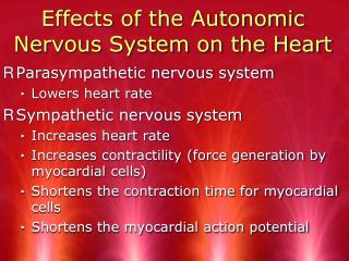
Cardiac sympathetic actions • Positive chronotropic effect正性变时作用 • Positive dromotropic effect正性变传导作用 • Positive inotropic effect正性变力作用
Mechanisms of norepinephrine —increase Na+ & Ca2+ permeability • If & ICa,T , phase 4 spontaneous depolarization, autorhythmicity • Ca2+ influx (ICa,L) , phase 0 amplitude & velocity , conductivity • Ca2+ influx (ICa,L) , Ca2+ release , [Ca2+ ]i , contractility (CICR)
Cardiac parasympathetic actions • Negative chronotropic effect负性变时作用 • Negative dromotropic effect负性变传导作用 • Negative inotropic effect负性变力作用
Mechanisms of acetylcholine • —increase K+ & decrease Ca2+ permeability • K+ outward (GIRK), If & ICa,T , |MRP|, phase 4 spontaneous depolarization , autorhythmicity • Inhibition of Ca2+ channel, phase 0 amplitude & velocity , conductivity • G protein→PLC → NOS → NO → GC → cGMP , Ca2+ influx (ICa,L) , [Ca2+ ]i , contractility
Vagal Maneuvers • Valsalva maneuver • A maneuver in which a person tries to exhale forcibly with a closed glottis (the windpipe) so that no air exits through the mouth or nose as, for example, in strenuous coughing, straining during a bowel movement, or lifting a heavy weight. The Valsalva maneuver impedes the return of venous blood to the heart. • Named for Antonio Maria Valsalva, a renowned Italian anatomist, pathologist, physician, and surgeon (1666-1723) who first described the maneuver.
Physiological response in Valsalva maneuver • The normal physiological response consists of 4 phases
Physiological response in Valsalva maneuver • The normal physiological response consists of 4 phases • Initial pressure rise: On application of expiratory force, pressure rises inside the chest forcing blood out of the pulmonary circulation into the left atrium. This causes a mild rise in stroke volume. • Reduced venous return and compensation: Return of systemic blood to the heart is impeded by the pressure inside the chest. The output of the heart is reduced and stroke volume falls. This occurs from 5 to about 14 seconds in the illustration. The fall in stroke volume reflexively causes blood vessels to constrict with some rise in pressure (15 to 20 seconds). This compensation can be quite marked with pressure returning to near or even above normal, but the cardiac output and blood flow to the body remains low. During this time the pulse rate increases. • Pressure release: The pressure on the chest is released, allowing the pulmonary vessels and the aorta to re-expand causing a further initial slight fall in stroke volume (20 to 23 seconds) due to decreased left ventricular return and increased aortic volume, respectively. Venous blood can once more enter the chest and the heart, cardiac output begins to increase. • Return of cardiac output: Blood return to the heart is enhanced by the effect of entry of blood which had been dammed back, causing a rapid increase in cardiac output (24 seconds on). The stroke volume usually rises above normal before returning to a normal level. With return of blood pressure, the pulse rate returns towards normal.
Tonus紧张 • Cardiac vagal tone心迷走紧张 • Cardiac sympathetic tone心交感紧张
Innervationof the blood vessels • Vasoconstrictor nerve缩血管神经 • Sympathetic vasoconstrictor nerve交感缩血管神经 • Vasodilator nerve舒血管神经 • Sympathetic vasodilator nerve交感舒血管神经 • Parasympathetic vasodilator nerve副交感舒血管神经 • Dorsal root vasodilator nerve脊髓背根舒血管神经
Cardiovascular Center A collection of functionally similar neurons that help to regulate HR, SV, and blood vessel tone
Vasomotor center Located bilaterally mainly in the reticular substance of the medulla and of the lower third of the pons • Vasoconstrictor area • Vasodilator area • Cardioinhibitor area – dorsal nuclei of the vagus nerves and ambiguous nucleus • Sensory area – tractus solitarius
Higher cardiovascular centers • Reticular substance of the pons • Mesencephalon • Diencephalon • Hypothalamus • Cerebral cortex • Cerebellum
Baroreceptor Reflexes压力感受性反射 • Arterial baroreceptors 动脉压力感受器 • Carotid sinus receptor • Aortic arch receptor • Afferent nerves (Buffer nerves,缓冲神经) • Cardiovascular center: medulla • Efferent nerves: cardiac sympathetic nerve, sympathetic constrictor nerve, vagus nerve • Effector: heart & blood vessels
Baroreceptor neurons function as sensors in the homeostatic maintenance of MAP by constantly monitoring pressure in the aortic arch and carotid sinuses.
Characteristics of baroreceptors: • Sensitive to stretching of the vessel walls • Proportional firing rate to increased stretching • Responding to pressures ranging from 60-180 mmHg • Receptors within the aortic arch are less sensitive than the carotid sinus receptors
The action potential frequency in baroreceptor neurons is represented here as being directly proportional to MAP.
i.e., MAP is above homeostatic set point i.e., reduce cardiac output Baroreceptor neurons deliver MAP information to the medulla oblongata’s cardiovascular control center (CVCC); the CVCC determines autonomic output to the heart.
Click here to play the Baroreceptor Reflex Control of Blood Pressure Flash Animation
Physiological Significance Maintaining relatively constant arterial pressure, reducing the variation in arterial pressure
Cardiovascular Responses to Exercise When exercise begins, mechanosensory input from working limbs combines with descending pathways from the motor cortex to activate the cardiovascular control center in the medulla oblongata. The center responds with sympathetic discharge that increases cardiac output and causes vasoconstriction in many peripheral arterioles.
Peripheral blood flow redistributes to muscle during exercise
Blood pressure rises slightly during exercise VO2 max (also maximal oxygen consumption, maximal oxygen uptake or aerobic capacity) is the maximum capacity of an individual's body to transport and utilize oxygen during incremental exercise, which reflects the physical fitness of the individual.
Baroreceptor reflex adjusts to exercise • During exercise, blood pressure increases without activating homeostatic compensation of baroreceptor reflex • Why? • Signal from the motor cortex during exercise reset the arterial baroreceptor threshold to a higher pressure