Download
1 / 11
130 likes | 785 Views
Regulation of stroke volume & heart rate. Measurement of cardiac output Regulation of heart rate neural Regulation of stroke volume Preload Afterload Neural Control of cardiac output. Measurement of cardiac output. Fick indicator-dilution method

E N D
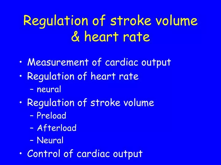
Regulation of stroke volume & heart rate • Measurement of cardiac output • Regulation of heart rate • neural • Regulation of stroke volume • Preload • Afterload • Neural • Control of cardiac output
Measurement of cardiac output • Fick indicator-dilution method • measures the time taken for an injected dye to pass a sampling point • Thermodilution • Echocardiography • Pulsed Doppler ultrasound
+25 0 mV -25 -50 -75 Regulation of heart rate • Sympathetic nervous system • sympathetic nerves release norepinephrine • plus circulating epinephrine from adrenal medulla • both act on ß-receptors on sinoatrial node • increases slope of the pacemaker potential • increases heart rate = tachycardia
+25 0 mV -25 -50 -75 Regulation of heart rate • Parasympathetic nervous system • vagus releases ACh • acts on muscarinic receptors on sinoatrial node • hyperpolarises cells and decreases slope of pacemaker potential • decreases heart rate = bradycardia
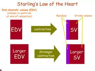
Tension Length Regulation of stroke volume - preload Starlings Law states - the energy of contraction is proportional to the initial length of the cardiac muscle fibre (= preload)
Stroke Volume Resting EDV End Diastolic Volume Regulation of stroke volume - preload In vivo, preload is affected by the End Diastolic Volume Increased venous return, increases EDV, and therefore increases stroke volume = self-regulation
Regulation of stroke volume - afterload • Afterload is the load against which the muscle tries to contract • In vivo, afterload is set by the arterial pressure against which the blood is expelled (this in turn depends on the Total Peripheral Resistance) • If TPR increases, stroke volume will go down
+ sympathetic stimulation Stroke Volume End Diastolic Volume Regulation of stroke volume - neural • Sympatheticnervous system • sympathetic nerves releasing norepinephrine • plus circulating epinephrine from adrenal medulla • both act on ß1-receptors on the myocytes • increases contractility (an inotropic effect) • gives stronger, but shorter contraction • Parasympathetic • little effect
Control of cardiac output HR x SV = CO • HR increases • via decrease vagal tone • & increased sympathetic tone • Contractility increases • via increased sympathetic tone • alters inotropic state & shortens systole • Venous return increases • via venoconstriction • & skeletal/respiratory pumps • maintains preload • Total peripheral resistance falls • due to arteriolar dilation in muscle, skin & heart • reduces afterload • CO increase 4-6 times
Summary • Heart rate • sympathetic supply HR • parasympathetic supply HR • Stroke volume • preload EDV, SV • afterload TPR, SV • neural sympathetic supply, SV • Think integration • these work together to produce a co-ordinated increase in CO, eg exercise