Download
1 / 35
360 likes | 617 Views
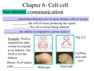
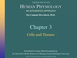
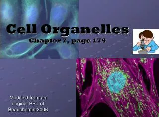
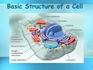
CHAPTER 7 – A VIEW OF THE CELL. Pg. 170 (not including section 7.2). 4. Lysosome. 5. Cytoskeleton. 6. Golgi apparatus. 3. Plasma membrane. 7. Centriole. 2. Cytoplasm. 8. Nucleus. 9. Nucleolus. 10. Mitochondria. 1. Vacuole. 11. E.R. 12. Ribosomes. 13. Animal Cell.

E N D
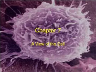
CHAPTER 7 –A VIEW OF THE CELL Pg. 170 (not including section 7.2)
4. Lysosome 5. Cytoskeleton 6. Golgi apparatus 3. Plasma membrane 7. Centriole 2. Cytoplasm 8. Nucleus 9. Nucleolus 10. Mitochondria 1. Vacuole 11. E.R. 12. Ribosomes 13. Animal Cell
THE HISTORY OF THE CELL THEORY Before microscopes, people believed diseases were caused by curses and spirits Cells can only be observed under a microscope Cells divide to create more cells There are two basic types of cells Eukaryotic Cells Prokaryotic Cells
Antonvan Leeuwenhoek Designed and built several hundred microscopes * Was the first to observe microscopic organisms The first to record observations of blood in capillaries Commonly known as the “Father of Microbiology”
Robert Hooke * Is credited with the discovery of the cell Studied cork, the dead cells of oak bark under a microscope Observed geometric shapes that he called “cells” because they reminded him of the small rooms at a monastery His observations drew some important conclusions by other scientists
Matthias Schleiden Observed plant structure under the microscope * Proposed that all plants are composed of cells Found fungi living on/in the roots
Theodor Schwann * Proposed that all living things are composed of cells Conducted experiments that disproved spontaneous generation Discovered the cells (Schwann cells) that surround nerve axons Discovered pepsin in the stomach of animals (and humans) Coined the term metabolism
Rudolph Virchow Concluded that the nucleus was responsible for cell division Is cited as the first to recognize leukemia * Proposed the theory that every cell comes from other cells
The Cell Theory The cell theory is made up of three ideas: 1. All organisms are composed of cells 2. The cell is the basic unit of structure and organization of organisms 3. All cells come from preexisting cells
Electron Microscopes Light microscopes can magnify up to 1500 times the actual size * Electron microscopes use a beam of electrons inside a vacuum Can magnify up to 500,000 times the actual size Scanning Electron Microscope (SEM) – Scans the surface of cells to get the 3D shape Transmission Electron Microscope (TEM) – Allows study structures within the cell (organelles) Scanning Tunneling Microscope (STM) – Uses the flow of electrons to create computer images of atoms on the surface of the conductive molecule Does anyone in the room have arachnophobia?
TWO BASIC CELL TYPES No matter which microscope you look through, all cells contain small, specialized structures called organelles However only the better microscopes allow us to observe organelles which have specific functions Prokaryotes (bacteria) are mostly unicellular organisms that do not have membrane-bound organelles not even a nucleus Eukaryotes are mostly multicellular that contain membrane-bound organelles Nucleus – Controls cellular function
CELLULAR BOUNDARIES Cell/plasma membrane = Flexible boundary made up of phospholipids, proteins and cholesterol surrounding the cell that allows the passage of materials into and out of the cell. Cell Wall – A fairly rigid structure found in plant cells and prokaryotic cells that is located outside of the plasma membrane for support and protection.
CELLULAR BOUNDARIES Cell/Plasma Membrane Structure
THE NUCLEUS AND CELL CONTROL Cytoplasm is the clear jelly-like watery fluid that holds the organelles outside the nucleus and gives the cell its shape. Also the site of many chemical reactions.
THE NUCLEUS AND CELL CONTROL The nucleus is bound by the nuclear membrane and contains the cell’s genetic material (DNA). It also acts as the control center for the cell and is responsible for cell division.
THE NUCLEUS AND CELL CONTROL Chromatin (“colored material”) – is the complex of DNA and proteins contained in the nucleus Condensed chromatin forms the chromosomes of each cell when it prepares to divide - X shaped DNA
THE NUCLEUS AND CELL CONTROL The nucleolus is the organelle within the nucleus that is surrounded by a condensed layer of chromatin The role of the nucleolus is to produce ribosomes
THE NUCLEUS AND CELL CONTROL Ribosomes - made up of proteins and RNA Produce all the proteins from DNA directions
THE NUCLEUS AND CELL CONTROL Nuclear envelope - surrounds nucleus and separates DNA (inside) from cytoplasm (outside) Nuclear pores - large protein complexes that allow for water-soluble molecules the pass through the nuclear envelope (NOT DNA)
ASSEMBLY, TRANSPORT, AND STORAGE The endoplasmic reticulum (ER) consists of a membrane network of tubes and sac-like structures It is located in the cytoplasm and is the site of chemical reactions
ASSEMBLY, TRANSPORT, AND STORAGE The rough ER- connected to the nuclear envelope and contains ribosomes which give it the “rough” appearance It works with the Golgi complex to transport new proteins to their proper destination via vesicles
ASSEMBLY, TRANSPORT, AND STORAGE The smooth ERdoes not contain ribosomes It is the site of lipid synthesis (fat creation) and carbohydrate metabolism It also houses key enzymes and enzyme reactions
ASSEMBLY, TRANSPORT, AND STORAGE The Golgi complex modifies, sorts, and packages proteins (from the ER) and lipids to be sent to the correct destination in vesicles (membrane bound compartments) It also transports lipids and is involved with lysosome creation
ASSEMBLY, TRANSPORT, AND STORAGE The vacuolesfound sometimes in animal cells are the membrane-bound compartments for temporary storage of materials (food, enzymes, waste) The vacuolefound in plant cells is a large “water tank” containing enzymes, ions, and salts Lysosomes contain digestive enzymes that destroy old organelles, food particles, and engulfed viruses or bacteria They can also fuse with vacuoles to digest their contents
ENERGY TRANSFORMERS Chloroplasts capture light energy in the stacks of thylakoids (grana) and convert it to chemical energy Contain chlorophyll which is the green pigment (protein) that traps light energy for photosynthesis
ENERGY TRANSFORMERS Mitochondria transform energy for the cell Membrane-bound organelle that have an outer membrane as well as a highly folded inner membrane Energy storing molecules are found on the inner membrane
SUPPORT & LOCOMOTION Cytoskeleton provides a constantly changing support structure Consists of thin, hollow cylinders made of proteins - microtubules Microfilaments are smaller, solid protein fibers that maintain the cell shape, anchor organelles, and provide a “material highway system”