Download
1 / 96
960 likes | 1.21k Views
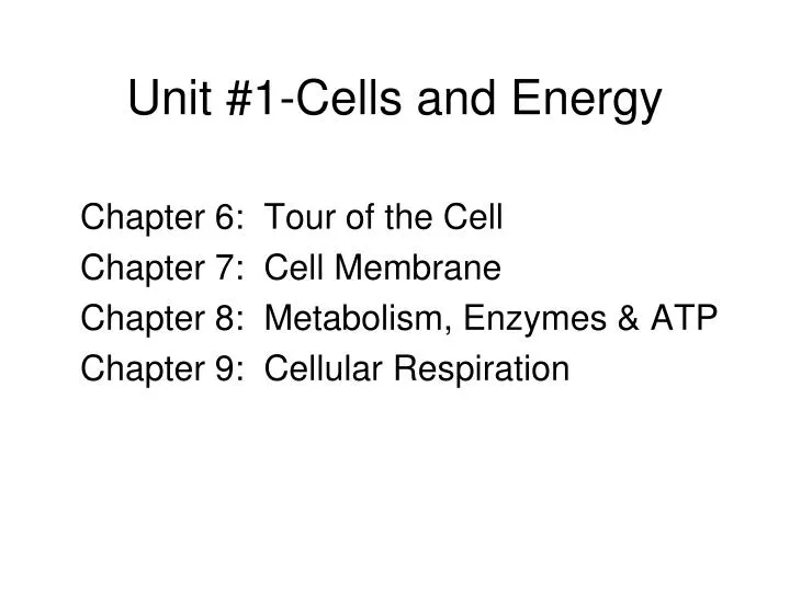
Unit #1-Cells and Energy. Chapter 6: Tour of the Cell Chapter 7: Cell Membrane Chapter 8: Metabolism, Enzymes & ATP Chapter 9: Cellular Respiration. A Tour of the Cell. Chapter 6 ( Pgs 94-123) History & discoveries Microscopy Limits to Cell Size (Surface area to volume ratio)

E N D
Unit #1-Cells and Energy Chapter 6: Tour of the Cell Chapter 7: Cell Membrane Chapter 8: Metabolism, Enzymes & ATP Chapter 9: Cellular Respiration
A Tour of the Cell Chapter 6 (Pgs 94-123) • History & discoveries • Microscopy • Limits to Cell Size (Surface area to volume ratio) • Cell Fractionation (Structure & Function of Organelles) • Prokaryotic vs.Eukaryotic • Plant cells vs. Animal • Endomembrane System • Cytoskeleton • Intercellular junctions
Video #1-Secrets of the Cell • What part of the football player’s body does the first segment focus on to show how cells work together? • What is Dr. Heath focusing on in plants? • Name three differences between prokaryotic and Eukaryotic cells mentioned in segment #3 of the video; **Write the title for each segment and FIVE key statements.
Below is a list of the most common units of length biologists use (metric) Table 4.2
Biological Size and Cell Diversity (Pg. 95) Human Eye: 1mm - meter+ LM: 1m – 1mm EM: 1nm – 1mm Chicken Egg (lgst cell) Mitochondria (1m) Ribosomes (20-30 nm) Viruses (80-100 nm)
History & Discovery of Cells • Anton Van Leeuwenhoek (1600’s) • Robert Hooke (Cork Cells, 1665) • Robert Brown (Nucleus, 1833) • Matthias Schleiden (Plant Cells, 1838) • Theodor Schwann (Animal Cells, 1839) • Rudolf Virchow (All Cells arise from other cells) • Cell Theory: 3 aspects
Microscopes provide windows to the world of the cell • The light microscope enables us to see the overall shape and structure of a cell Image seen by viewer Eyepiece Ocularlens Objective lens Specimen Condenser lens Light source Figure 4.1A
Scanning electron microscope (SEM) SEM of cilia View SEM Images: http://micro.magnet.fsu.edu/primer/java/electronmicroscopy/magnify1/index.html Figure 4.1B
Transmission electron microscope (TEM) • Transmission electron micrograph of cilia http://www.lifesci.sussex.ac.uk/home/Julian_Thorpe/TEM3.htm Figure 4.1C
Cytology: science/study of cells • Light microscopy •resolving power~ measure of clarity • Electron microscopy (2 types) •TEM~ electron beam to study cell ultrastructure •SEM~ electron beam to study cell surfaces • Cell fractionation~ cell separation; organelle study • Ultracentrifuges~ cell fractionation; 130,000 rpm
Cell Fractionation • Physically separates and purifies cell parts • Spun in a centrifuge (up to 500,000 rpm) • Two fractions: supernatant & pellet • Differential: successively at higher speeds • Density gradient: forms bands in tube according to density differences of organelles
Cell Size • Is it more advantageous to be a single cell that is large or to be broken down into several small cells ? (Explain your answer)
Natural laws limit cell size • At minimum, a cell must be large enough to house the parts it needs to survive and reproduce • The maximum size of a cell is limited by the amount of surface needed to obtain nutrients from the environment and dispose of wastes
A small cell has a greater ratio of surface area to volume than a large cell of the same shape 30 µm 10 µm Surface areaof one large cube= 5,400 µm2 Total surface areaof 27 small cubes= 16,200 µm2 Figure 4.3
Cell size - (surface area:volume) • As cell size increases, the surface area to volume ratio decreases (sa/vol) • Rates of chemical exchange may then be inadequate for cell size • Cell size, therefore, remains small
A prokaryotic cell is enclosed by a plasma membrane and is usually encased in a rigid cell wall • The cell wall may be covered by a sticky capsule Prokaryoticflagella Ribosomes Capsule Cell wall • Inside the cell are its DNA and other parts Plasma membrane Nucleoid region(DNA) Pili Figure 4.4
Prokaryotic cells, Bacillus polymyxa Figure 4.4x1
Prokaryotic cell, E. coli Figure 4.4x2
Pili on a prokaryotic cell Figure 4.4x3
Prokaryotic flagella Figure 4.4x4
The Prokaryotic Cell-(See Fig. pg 98)(Also See Pages534-547 in Ch. 27) • Characteristics include: • No true distinct nucleus • Have a “Nucleoid” region = DNA & Plasmids • No complex, membranous organelles (Ribosomes only) • Most have cell walls • Flagella (rotary type structure & not composed w/microtubules) • Some have pigments (autotrophic) • Classified according to their metabolic needs • Eubacteria & Archeabacteria • Some have Capsules, pili, peptidoglycan, Endospores • Asexually Reproduce: Binary Fission, Budding, Fragmentation • Genetic Material Can be exchanged by 3 mechanisms: • Transformation, Transduction, and Conjugation
Prokaryotic and eukaryotic cells compared Figure 4.4x5
The Eukaryotic Cell • “Eu” = true “Karyo” = kernal (nucleus) • Protists, Plants, Fungi, and Animals • Internal Membrane System • Has many membranous organelles (Table 4.1) that include: -Nucleus -Lysosomes -Golgi complex -Endoplasmic reticulum (R & S) -Mitochondria -Chloroplast (plastids) -Peroxisomes (glyoxysomes) -Vesicles -Vacuole (food, contractile)-Ribosomes • Cytoskeleton: microtubules, microfilaments, and int. filaments • Centrioles (nine triplets of microtubules) • Cilia & Flagella (9+2 microtubule arrangement) • Extracellular matrix (ECM)-proteins & carbodydrate -glycoproteins -glycolipids -integrins -fibronectins -collagen
Roughendoplasmicreticulum Nucleus Ribosomes Smoothendoplasmicreticulum Golgiapparatus Microtubule Centralvacuole Not inanimalcells Intermediatefilament Cytoskeleton Chloroplast Microfilament Cell wall Mitochondrion Peroxisome Plasma membrane Figure 4.5B
Plant Cell • Cell wall • Chloroplasts • Water Vacuole • Mitochondria
Smooth endoplasmicreticulum Nucleus Roughendoplasmicreticulum • An animal cell Flagellum Not in most plant cells Lysosome Centriole Ribosomes Peroxisome Golgiapparatus Microtubule Plasma membrane Cytoskeleton Intermediatefilament Microfilament Mitochondrion Figure 4.5A
Animal Cell • Centrioles • Mitochondria • Plasma Membrane
Nucleus, Ribosomes, Rough & Smooth ER, Flow of Genetic information and protein Synthesis
Nucleus (Pg. 103) Control Center of the Cell Genetic material: •chromatin •chromosomes Nucleolus: ribosome synthesis Double membrane envelope with pores 1st part of Protein synthesis: Transcription (DNAmRNA) Nuclear pores
NUCLEUS Chromatin Two membranesof nuclearenvelope Nucleolus Pore ROUGHENDOPLASMICRETICULUM Ribosomes Figure 4.6
Ribosomes • Manufactures Protein • Free •cytosol; •protein function in cell • Bound •endoplasmic reticulum; •membranes, organelles, and export
Endoplasmic Reticulum (pg. 105) Endoplasmic reticulum (ER) • Continuous with nuclear envelope Smooth ER •no ribosomes •synthesis of lipids, hormones, and steroids ***Abundant in testes, ovary, and adrenal glands •Metabolism of carbohydrates •Detoxification of drugs and poisons (Liver) Stores calcium ions (muscle cells---sarcoplasmic reticulum) Rough ER •with ribosomes •synthesis of secretory proteins (glycoproteins), membrane production **Found extensively in Pancreas & nerve cells
SMOOTH ER ROUGHER Nuclearenvelope Ribosomes SMOOTH ER ROUGH ER Figure 4.9
Transport vesiclebuds off 4 Ribosome Secretory(glyco-) proteininside transportvesicle Sugarchain 3 Glycoprotein 1 2 ROUGH ER Polypeptide Rough Endoplasmic Reticulum makes membrane and proteins • The rough ER manufactures membranes • Ribosomes on its surface produce proteins Figure 4.8
Golgi Complex (pg. 106) • Golgi apparatus • •ER products are modified, stored, and then shipped • Cisternae: flattened membranous sacs • trans face (shipping) & cis face (receiving) • Transport vesicles
The Golgi apparatus finishes, sorts, and ships cell products • The Golgi apparatus consists of stacks of membranous sacs • These receive and modify ER products, then send them on to other organelles or to the cell membrane • Specialized for secretion (salivary glands & pancreas) • Removes and changes the sugars attached to the protein • Many polysaccharides are secreted by the Golgi
The Golgi apparatus Golgi apparatus Golgiapparatus “Receiving” side ofGolgi apparatus Transportvesiclefrom ER Newvesicleforming “Shipping”side of Golgiapparatus Transport vesiclefrom the Golgi Figure 4.10
Lysosomes digest the cell’s food and wastes (Pg.107) • Lysosomes are sacs of digestive enzymes budded off the Golgi LYSOSOME Nucleus Figure 4.11A
Contain lysosomal enzymes (hydrolytic enzymes) • digests food molecules (macromolecules) • destroys bacteria • recycles damaged organelles • function in embryonic development in animals • undergoes phagocytosis & engulfs material • Recycle cell’s own organic material • **Found extensively in Macrophages (WBC’s) Lysosomes:
Rough ER Transport vesicle(containing inactivehydrolytic enzymes) Plasmamembrane Golgiapparatus Engulfmentof particle Lysosomeengulfingdamagedorganelle “Food” LYSOSOMES Digestion Foodvacuole Figure 4.11B
Lysosomes can cause Fatal Diseases • Lysosomal Storage Diseases are hereditary that interfere with other cellular functions *Examples: Pompe’s disease Tay-Sachs disease (Pgs. 93, 331)
Vacuoles -Membrane-bound sacs (larger than vesicles) -Food (phagocytosis) -Contractile (pump excess water) -Central (storage in plants) -Tonoplast membrane
Vacuoles function in the general maintenance of the cell • Plant cells contain a large central vacuole • The vacuole has lysosomal and storage functions Centralvacuole Nucleus Figure 4.13A
Peroxisomes (Pg. 111) • Single membrane • Oxidative organelle ***strips e-’s (H’s) from substances • Produce hydrogen peroxide (H2O2) in cells • Metabolism of fatty acids; detoxification of alcohol (liver) • Hydrogen peroxide then converted to water