Download

1 / 72
770 likes | 1.36k Views
Lecture 4: Chapter 4 The Tissue Level of Organization Pages: 106 - 152. Lecturer: Dr. Barjis Room: P313 Phone: (718) 260-5285 E-Mail: ibarjis@citytech.cuny.edu. Learning Objectives. Identify the four major tissue types and describe their functions.

E N D
Lecture 4: Chapter 4The Tissue Level of OrganizationPages: 106 - 152 Lecturer: Dr. Barjis Room: P313 Phone: (718) 260-5285 E-Mail: ibarjis@citytech.cuny.edu
Learning Objectives • Identify the four major tissue types and describe their functions. • Describe the relationship between form and function for each tissue type. • Discuss the types and functions of epithelial tissues. • Compare the structure and function of connective tissues.
Learning Objectives • Explain the structure and function of the four types of membrane. • Describe the three types of muscle tissue and the structural features of each. • Discuss the basic structure and role of neural tissue.
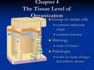
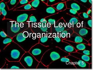
Tissues of the Body: An Introduction Tissues and tissue types • Tissues are: • Collections of specialized cells and cell products organized to perform a limited number of functions • Histology = study of tissues • The four tissue types are: • Epithelial • Connective • Muscular • Nervous
Tissues and tissue types Epithelial tissue • Includes glands and epithelium • Glands are secretory • Is avascular • Forms a protective barrier that regulates permeability • Cells may show polarity
Functions of epithelium • Physical protection • Control permeability • Provide sensation • Produce specialized secretions
Specializations of epithelium • Perform secretory functions • Perform transport functions • Maintain physical integrity • Ciliated epithelia move materials across their surface
Maintaining the integrity of epithelium • Cells attach via cell adhesion molecules (CAM) • Cells attach at specialized cell junctions • Tight junctions • Desmosomes • Gap junctions
Intercellular connections Animation: check tutorials
Structure of typical epithelium • Basal lamina attaches to underlying surface • Lamina lucida • Lamina densa • Germinative cells replace short-lived epithelial cells
Classification of epithelia • Number of cell layers • Simple • Stratified • Shape of apical surface cells • Squamous • Cuboidal • Columnar
Glandular epithelia • Exocrine glands • Secrete through ducts onto the surface of the gland • Endocrine glands • Release hormones into surrounding fluid
Glandular secretions can be: • Merocrine (product released through exocytosis) • Apocrine (involves the loss of both product and cytoplasm) • Holocrine (destroys the cell)
Mechanisms of Glandular Secretion Animation: Mechanisms of glandular secretion(check tutorial)
Glands • Unicellular • Individual secretory cells • Multicellular • Organs containing glandular epithelium • Classified according to structure
Connective Tissues Connective tissue functions: • Establishing a structural framework • Transporting fluids and dissolved materials • Protecting delicate organs • Supporting, surrounding and interconnecting tissues • Storing energy reserves • Defending the body from microorganisms
Connective tissues contain • Specialized cells • Matrix • Composed of extracellular protein fibers and a ground substance
Connective tissue proper • Contains varied cell populations • Contains various fiber types • A syrupy ground substance
Fluid connective tissue • Contains a distinctive cell population • Watery ground substance with dissolved proteins • Two types • Blood • Lymph
Supporting connective tissues • Less diverse cell population • Dense ground substance • Closely packed fibers • Two types • Cartilage • Bone
Connective tissue proper • Contains fibers, a viscous ground substance, and a varied cell population • Fibroblasts • Macrophage • Adipocytes • Mesenchymal cells • Melanocytes • Mast cells • Lymphocytes • Microphages
Connective tissue proper • Three types of fiber • Collagen fibers • Reticular fibers • Elastic fibers
Connective tissue proper • Classified as loose or dense • Loose • Embryonic mesenchyme, mucous connective tissues • Areolar tissue • Adipose tissue • Reticular tissue • Dense • Dense regular CT • Dense irregular CT
Fluid connective tissues • Distinctive collections of cells in a fluid matrix • Blood • Formed elements and plasma • Red blood cells, white blood cells and platelets • Arteries carry blood away, veins carry to the heart • Capillaries allow diffusion into the interstitial fluid • Lymph • Interstitial fluid entering the lymphatic vessels
Supporting connective tissues • Cartilage and bone support the rest of the body • Cartilage • Grows via interstitial and appositional growth • Matrix is a firm gel containing chondroitin sulfate • Cells called chondrocytes • Cells found in lacunae • Perichondrium separates cartilage from surrounding tissues • Three types: hyaline, elastic and fibrocartilage
Bone, or osseus tissue • Has osteocytes • Depend on diffusion through canaliculi for nutrients • Little ground substance • Dense mineralized matrix • Surrounded by periosteum
Membranes Membranes are simple organs • Form a barrier • Composed of epithelium and connective tissue • Four types • Cutaneous • Synovial • Serous • Mucous
Mucous membranes • Line cavities that communicate with the exterior • Contain lamina propria