Download
1 / 78
800 likes | 1.07k Views
Adrenal Disease. Jennii Stephens, PA-C. Diseases of the Adrenal Gland. Anatomy and Physiology Decreased adrenal function Cortex Addisons Disease Secondary hypoadrenalism (pituitary dysfunction) Increased adrenal function Cortex: Cushings Syndrome/Disease Conn Syndrome

E N D
Adrenal Disease Jennii Stephens, PA-C
Diseases of the Adrenal Gland • Anatomy and Physiology • Decreased adrenal function • Cortex • Addisons Disease • Secondary hypoadrenalism (pituitary dysfunction) • Increased adrenal function • Cortex: • Cushings Syndrome/Disease • Conn Syndrome • Medulla: Pheochromocytoma
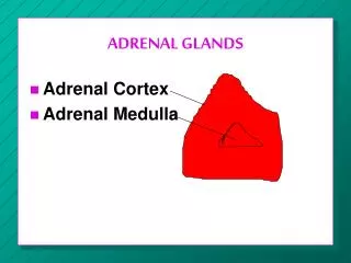
Adrenal Anatomy “salt” “sugar” “sex” “GFR”—salt, sugar, sex, the deeper you go the sweeter it gets
Adrenal Cortex Layers • Glomerulosa • Mineralocorticoids, aldosterone • maintains sodium and potassium water balance via the distal tubules of the kidney • Fasciculata • Glucocorticoids, cortisol • regulates carbohydrate, protein and fat metabolism
Adrenal Cortex Layers • Reticularis • Androgens and estrogens • These are produced in far greater abundance in gonads • No other tissues have the capability of producing either mineralocorticoids or glucocorticoids
Adrenal medulla • Inner portion of adrenal gland • Secretes epinephrine and norepinephrine • Acts to reinforce activity of the sympathetic nervous system • Not vital to life (its absence does NOT cause disease)
Mineralocorticoids • Aldosterone • Site of action is distal tubules of kidney • Retain Nablood volumeblood pressure • Crucial for sodium conservation in • Kidney • Salivary glands • Sweat glands • Colon • Absolutely essential for life
Aldosterone regulation BP Direct renin inhibitor (Tekturna, aliskiren) Renin Angiotensinogen Angiotensin I ACE inhibitors (‘pril’: lisinipril, captopril…) ACE Angiotensin II Angiotensin receptor blocker (ARB), (‘sartan’: valsartan, losartan…) ALDOSTERONE Na reabsorption Blood volume BP
Glucocorticoids (cortisol) • blood glucose • hepatic gluconeogenesis • glucose uptake and use by many tissues, but not the brain • protein degradation which frees amino acids for gluconeogenesis • lipolysis, releasing fatty acids which can act as an alternative metabolic fuel for tissues • Frees glucose for brain usage • Anti-inflammatory and immunosuppressant • Synthetic forms maximize these characteristics allowing us to use them clinically
Glucocorticoids (cont) • Secretion of cortisol is controlled by ACTH from the anterior pituitary • pro-opiomelanocortin is broken down to form ACTH and melanocyte-stimulating hormone (MSH) • Thus, high ACTH means high MSHincreased skin pigmentation
Androgens • Produced by the zona reticularis • Primary area of androgen production in women
Diseases of the Adrenal Gland • Anatomy and Physiology • Decreased adrenal function • Cortex • Addisons Disease • Secondary hypoadrenalism (pituitary dysfunction) • Increased adrenal function • Cortex: • Cushings Syndrome/Disease • Conn Syndrome • Medulla: Pheochromocytoma
Primary v Secondary • Primary disease • Pathology at the organ • Secondary disease • Pathology somewhere other than the organ • The organ has the ability to function normally
Addison’s • 1-4 in 100,000 people • Most common in adults 30-50 yo • Primaryhypoadrenalism • Pay attention to the distinction between primary and secondary characteristics
Pathophysiology of Addisons • Due to destruction of >90% of bilateral adrenal cortices (80% of cases) • Types • Usually autoimmune • Can be associated with other autoimmune diseases (Graves, type I DM, pernicious anemia…) • Takes months to years to destroy this much cortex • Thrombosis • Hemorrhage • Infectious causes (HIV, Tb, fungal…) • Cancer • Certain drugs • Affects both glucocorticoid and mineralocorticoid function
Pathophysiology of Secondaryhypoadrenalism • Due to lack of ACTH • Results in deficiency of cortisol • Thus, aldosterone is NOT affected • Causes • Pituitary disease, such as tumor • Prolonged use of steroid medication is discontinued without appropriate taper • Infection • Head trauma • Pituitary infarction (Sheehan’s syndrome)
Effects of low aldosteroneAddison’s disease • Na excretion • K secretion • Thus, get rid of Naget rid of watervolume depletionlow BP
Effects ofcortisol deficiencyAddison’s disease or Secondary hypoadrenalism • Carbohydrate metabolism disturbed • Hypoglycemiaweakness • Addison’s disease • ACTH is increased in response to low cortisol • This stimulates MSH (melanocyte stimulating hormone)hyperpigmentation
Chronic presentation • Hyperpigmentation of skin and mucous membranes • Caused by ACTH stimulatory effect on melanocytes • Most prominent on • sun-exposed areas • Knuckles, elbows, knees and new scars • Palmar creases, nail beds, oral cavity, vaginal and perianal mucosa
Hyperpigmented fingers and nails Fingers of a 28-year-old white woman with Addison's disease (underneath) compared to those of a normal woman (top). There is hyperpigmentation of the skin and increased pigmentation of the distal half of the nails that occurred during the period of adrenal insufficiency. The proximal half of the nails are hypopigmented, a reflection of the reduction in ACTH secretion after the institution of glucocorticoid therapy. Courtesy of David N Orth, MD.
Chronic presentation • Progressive weakness • Chronic, worsening fatigue • Poor appetite, craving of salty foods • Weight loss • Hypotension (Addison’s) • GI symptoms (n/v/d) • Dizziness (orthostatic hypotension) • Irritability and depression • Myalgia and flaccid muscle paralysis (from hyperkalemia) decreased/absent reflexes • Addison’s disease only
Chronic presentation • Men • Impotence • Decreased libido • Women • Decreased body hair (from decreased androgens) • Irregular menses or amenorrhea • Due to chronic ill health/ weight loss/ autoimmune destruction of ovarian tissue
Lab Studies • BMP • Hyponatremia, Hyperkalemia, Mild non-anion gap metabolic acidosis (Addison’s only) • due to lack of sodium-retaining and potassium and hydrogen ion-secreting action of aldosterone • Elevated BUN and creatinine(Addison’s only) • due to hypovolemia, a decreased GFR, and decreased renal plasma flow • Hypercalcemia (unknown mechanism) • Hypoglycemia (Both primary and secondary dz) • Caused by increased peripheral utilization of glucose and increased insulin sensitivity • Urinary and sweat sodium elevated (Addison’s only)
Diagnosis of Adrenal Insufficiency • ACTH stimulation test (Cortrosyn) • Assesses functional capacity of adrenal glands to make cortisol • Draw cortisol level • Inject ACTH • Wait 30-60 minutes • Draw cortisol levels • If normal adrenal functionhigher cortisol • If abnormal adrenal functionno change • If secondary diseasecan be no change to higher levels depending on the chronicity of the disease
Imaging Studies • CT—depends on cause • Infectious—will often show enlarged adrenals • TB, histoplasmosis—calcification of adrenals • Autoimmune—atrophic adrenals
Other studies • EKG—changes due to hyperkalemia
Acute presentationAddisonian Crisis • Prominent n/v • Vascular collapse (shock) • Confused • Cyanotic • Hyperpyrexia (may reach 105ºF) • Abdominal symptoms may appear like an acute abdomen
Acute causes • Stress (infection, trauma, surgery, emotional turmoil) with failure to increase steroids in patient with chronic Addison’s • Abrupt cessation of chronic oral steroids • COPD patient • Bilateral adrenal hemorrhage • Fulminant meningococcemia • Bilateral adrenal artery emboli • Sepsis, DIC • Medications: rifampin, ketoconazole, phenytoin
Treatment of Acute Adrenal Crisis • Begin immediate treatment with salt, fluids and glucocorticoids • draw random plasma cortisol level (before any glucocorticoids given)
Treatment • Replace cortisol • Acute adrenal crisis: IV • Clinical improvement (BP response) should be seen within 4-6h of tx • Taper dose after response • Chronic • Daily oral replacement of both mineralocorticoid and corticosteroid • For lifetime (can’t ever stop) • Will have to increase doses during periods of stress
Chronic meds • Hydrocortisone • Drug of choice for acute and chronic tx in Addison’s • Has both glucocorticoid and mineralocorticoid properties • Titrate to patient’s general well being and presence of symptoms • Patients should be told to double or triple their steroid replacement doses in stressful situations (common cold, tooth extraction…)
Chronic Meds (cont) • Fludrocortisone • Very potent mineralocorticoid • Oral med • Titrate to maintain normal BP, Na, and K levels • No dose adjustment in stressful situations
Miscellaneous • Should consult endocrinologist • Patient should wear medical ID bracelet • Prognosis: with adequate therapy, normal lives
Diseases of the Adrenal Gland • Anatomy and Physiology • Decreased adrenal function • Cortex • Addisons Disease • Secondary hypoadrenalism (pituitary dysfunction) • Increased adrenal function • Cortex: • Cushings Syndrome/Disease • Conn Syndrome • Medulla: Pheochromocytoma
Cushings • 13 per million patients • Usually due to exogenous glucocorticoids • 5 women:1 man • Usually 25-40 yo • Two distinctions: • Syndrome--A group of conditions caused by increased production of cortisol hormones or by the administration of glucocorticoid hormones • Disease—a form of Cushing’s syndrome caused by an ACTH-secreting pituitary tumor
Presentation • Increased adipose tissue • moon facies • buffalo hump (on upper back at base of neck) • supraclavicular fat pads • truncal obesity • facial plethora(flushed) • purple striae • usually >1cm in width • Most commonly abdomen, buttocks, lower back, upper thighs and arms, breasts
Moon Facies 30-year-old woman with Cushing's disease showing round, plethoric "moon" face, facial hirsutism, and increased supraclavicular fat pads. Williams Textbook of Endocrinology, 8th ed, Foster, DW, Wilson, JD (Eds), WB Saunders, Philadelphia, 1996.