Download
1 / 17
170 likes | 389 Views
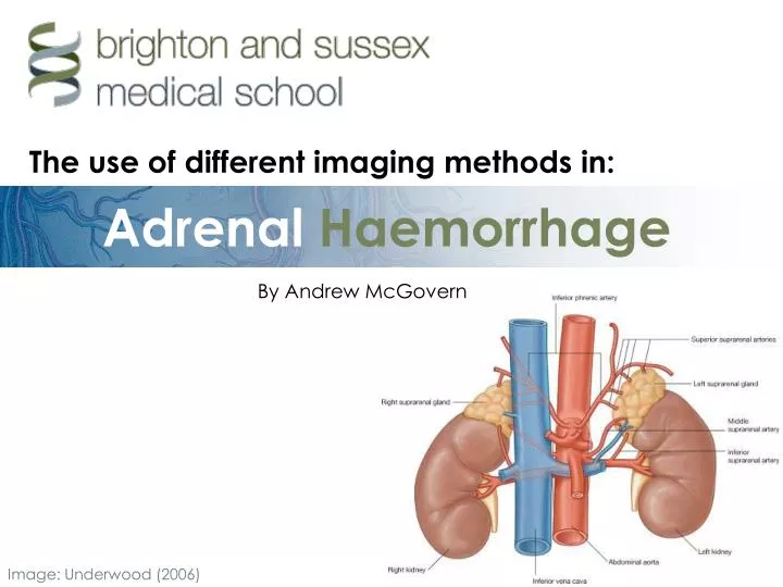
Adrenal Haemorrhage. The use of different imaging methods in:. By Andrew McGovern. Image: Underwood (2006). Introduction. PART 1. Adrenal haemorrhage: An overview Pathophysiology PART 2. Imaging in adrenal haemorrhage: Different imaging possibilities

E N D
AdrenalHaemorrhage The use of different imaging methods in: By Andrew McGovern Image: Underwood (2006)
Introduction PART 1. Adrenal haemorrhage: • An overview • Pathophysiology PART 2. Imaging in adrenal haemorrhage: • Different imaging possibilities • Positive and negative aspects of each type • Image examples
Adrenal haemorrhage at autopsy Image: Underwood (2004) What is adrenalhaemorrhage? • Uncommon, usually presents as bilateral haemorrhage. (Light, 2006) • Can occur at any age. (Light, 2006) • Extensive necrosis of all three cortical layers and medulla. (Tritos, 2007) • Can cause volume lossand shock in infants and acute adrenalinsufficiency unlessrecognised andtreated properly.(Light, 2006)
Pathophysiology Causes of adrenal haemorrhage: • Coagulopathies: causing thrombosis in renal and adrenal veins. • Waterhouse-Friderichsen syndrome: in children and young adults (occurring in 20% of meningitis cases). • Trauma: found in 28% of severe trauma cases autopsy. (Sevitt, 1955) • Asphyxia: in neonates – at birth the adrenal gland is very large and vascular. • Also associated with adrenal tumours. (Light, 2006)
Imaging:Computed tomography CT scanning is the preferred method for identifying adrenal haemorrhage in all patients over 6 months old. CT is rapid, widely available and accurate in diagnosis. • Useful for the identification of an underlying neoplasm, tumour or large thrombosis. • Allows examination of the adrenal glands in trauma patients with other imaging indications. Adrenal haemorrhage is detected as a round or oval mass obliterating the normal chevron shape of the adrenal gland. (Light, 2006)
Imaging:Computed tomography CT of normal adrenals several months before the onset of haemorrhage. CT two weeks after the onset of an acute haemorrhage. Images excerpted from: Rao et al. (1989)
Imaging:Computed tomography CT of acute bilateral haemorrhage. Image: Hentel (2008)
Imaging:Magnetic Resonance In general MRI identifies adrenal abnormalities at a rate which is comparable to CT. • Not as widely available as CT. • May be preferable in younger people due to CT radiation risk: • 1 fatal cancer per 2000 scans. (FDA, 2007) • 20% increased lifetime cancer mortality risk. (FDA, 2007) • 5 fatal cancers per 6000 scans in under 15s. • May account for up to 2% of cancers in the US.(Brenner, 2007) • Evidence for these studies is 25,000 survivors of Hiroshima bombing. Not directly comparable?(BRER, 2006)
Imaging:Magnetic Resonance T1 weighted MRI displaying right adrenal infarction without haemorrhage, in a 42-year-old man with anti phospholipid syndrome. Image: Riddell and Khalili (2004)
Imaging:Ultrasound US is the standard in neonates for imaging adrenal masses. • It is sensitive for enlargements of the adrenal glands. • Can differentiate between causes of adrenal mass. • Echogenicity is variable with the age of the haemotoma – can estimate age. • Widely available. • No ionising radiation. • But US is operator dependant. (Light, 2006)
Imaging:Ultrasound An adrenal haemorrhage as seen on ultrasound. Image: Hentel (2008)
Imaging:Ultrasound An adrenal haemorrhage as seen with Doppler ultrasound. This enables the avascular nature of the mass to be identified. Image: Hentel (2008)
Imaging:Ultrasound Adrenal haemorrhage in a foetus at 34 weeks gestation caused by a neuroblastoma. Image: Trop and Levine (2001)
Imaging:Other modalities Plain radiographs: Acute adrenal haemorrhage is rarely detectable. It may cause mass effect in the upper abdomen. (Light, 2006) Image: Kawashima et al. (1999)
Imaging:Other modalities Nuclear medicine studies:Of very little use as the main contrast agents used are not taken up by haematoma or normal adrenal tissue. (Light, 2006) Image: Kawashima et al. (1999)
Summary • Adrenal haemorrhage is an uncommon but serious condition which causes adrenal insufficiency. • CT is the diagnostic method of choice. • MRI should be considered as an alternative in children. • Ultrasound should be used in neonates.
References BRENNER, D. J. (2007) Computed Tomography — An Increasing Source of Radiation Exposure. the New England Journal of Medicine,357 pp. 2277-2284. BRER (2006) Health risks from exposure to low levels of ionizing radiation — BEIR VII, 1st ed. Washington, DC: The National Academies Press. FDA (2007) What are the Radiation Risks from CT? [online]. Available from: http://www.fda.gov/cdrh/ct/risks.html [accessed: 28 Feb 2008]. HENTEL, B. (2008) Adrenal hemorrhage [online]. Available from: http://www.radswiki.net/main/index.php?title=Adrenal_hemorrhage [accessed: 23 Feb 08]. KAWASHIMA, A., SANDLER, C. M., ERNST, R. D., TAKAHASHI, N., ROUBIDOUX, M. A., GOLDMAN, S. M. et al. (1999) Imaging of Nontraumatic Hemorrhage of the Adrenal Gland. RadioGraphics,19 pp. 949-963. LIGHT, D. (2006) Adrenal Hemorrhage [online]. Available from: http://www.emedicine.com/radio/topic15.htm [accessed: 23 Feb 2008]. RAO, R. H., VAGNUCCI, A. H. and AMICO, J. A. (1989) Bilateral massive adrenal hemorrhage: early recognition and treatment. Annals of Internal Medicine,110 (3), pp. 227-235. RIDDELL, A. M. and KHALILI, K. (2004) Sequential Adrenal Infarction Without MRI-Detectable Hemorrhage in Primary Antiphospholipid-Antibody Syndrome. American Journal of Roentgenology,183 pp. 220-222. SEWITT, S. (1995) Post-traumatic adrenal apoplexy. Journal of Clinical Pathology,8 (3), pp. 184-194. TRITOS, N. A. (2007) Adrenal Hemorrhage [online]. Available from: http://www.emedicine.com/MED/topic3009.htm [accessed: 28 Feb 2008]. TROP, I. and LEVINE, D. (2001) Hemorrhage During Pregnancy Sonography and MR Imaging. American Journal of Roentgenology,176 pp. 607-615. UNDERWOOD, J. C. E. (Ed.) (2004) General and Systemic Pathology, 4th ed. London: Churchill Livingstone.