Download
1 / 14
180 likes | 666 Views
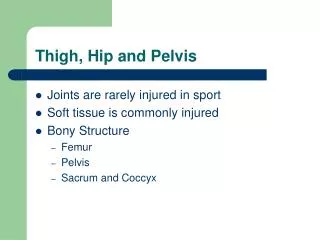
Hip and Pelvis. Muscle Tests. Gaenslen’s Sign. Used to assess if pathology exists in sacroiliac joint Patient lies supine on table with both knees bend and hug knees to chest Patient located at end of table to one buttock is over edge. Gaenslen’s Sign.

E N D
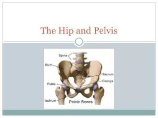
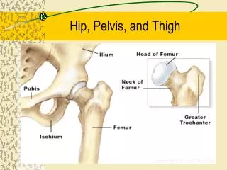
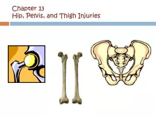
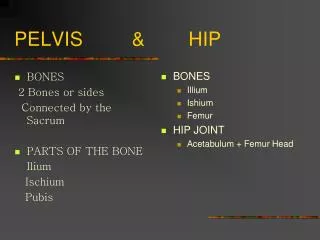
Hip and Pelvis Muscle Tests
Gaenslen’s Sign • Used to assess if pathology exists in sacroiliac joint • Patient lies supine on table with both knees bend and hug knees to chest • Patient located at end of table to one buttock is over edge
Gaenslen’s Sign • Both shoulders remain on table as long as full range of hip extension is permitted • Therapist stabilizes patient at torso so patient feels balanced • Keeping supporting leg flexed, allow unsupported leg to drop over edge of table • Ask if any pain • Pain in area of sacroiliac joint may indicate pathology • Test both sides
Thomas Test • Used to help detect flexion contractures of the hip or to evaluate range of hip extension • Muscle contracture: a muscle that fails to relax completely • Orthopedic contractures: shortening of the connective tissue (Houglam, 2005)
Thomas Test • Patient lies supine on table • Ensure pelvis is level and square to trunk • Stabilize the pelvis by placing hand under lumbar spine • Bend patient’s knee to chest, flex hip • Therapist notes the point that the back flattens, at this point the movement is purely hip joint and no pelvic movement • Flex hip as far as possible
Thomas Test • Thigh should rest against abdomen • Flex other hip • Patient holds one leg and extend the other as far as possible • Leg should rest flat on table • If leg does not lie flat, may indicated a fixed flexion contracture • Rocking forwards or lifting of the thoracic spine and return of lumbar lordosis are indicators of contracture • Approximate angle between the extended leg and the table
Trendelenburg Test • Tests the stability of the hip and evaluate the strength of the gluteus medius because it is the main stabilizer (Adams, 1971) • Patient stands erect bearing weight evenly on both feet • Therapist stands behind the patient • Note where posterior superior iliac spines are, should be level
Trendelenburg Test • Therapist asks patient to stand on one leg • Gluteus medius of support leg should contract and elevate hip of unsupported side (negative result) • If there is a weakness or gluteus medius is not functioning, the hip of the unsupported side will remain level or may drop below level of supported side (positive result)
Test for Leg Length Discrepancy • To determine the general leg length discrepancy • Patient lies supine and therapist ensures that patient is lying straight with no curves in spine or pelvic tilt • Therapist measures from the superior iliac spine to medial malleoli
Test for Leg Length Discrepancy • If measurements are even, no leg length discrepancy • If there is a difference, testing should be continued to determine where discrepancy exists
Tibial or Femoral Discrepancy • Flex knees to ninety degrees • Tibial discrepancy: standing in front of patients knees, therapist will notice one of the knees higher than the other • Femoral discrepancy: standing lateral to patient, therapist will notice one leg projecting past the other anteriorly
Ober Test • Test of the iliotibial band • Testing for contracture of the band or the fascia lata • Patient lies on side with involved side up • Keeping hip neutral the therapist supports medial side of distal femur and medial side of tibia
Ober Test • Therapist abducts leg at hip as much as possible then flexes knee to 90º • Therapist slowly releases leg down • If leg drops to adducted position no contracture exists (negative Ober) • If leg remains abducted, contractureexists (positive ober)
References • Adams, J.C.(1971). Outline of orthopaedics (7th ed.). E.&S. Livingstone Limited: London. • Hoppenfield, S. (1972). Physical examination of spine and extremities. Applteton Croft. • Houghlum, P.A. (2005). Therapeutic exercises for musculoskeletal injuries. Human Kinetics.