Download
1 / 65
750 likes | 862 Views
Management of Thyroid Cancer. Local seminar Medical Oncology department. By Salah Mabruok Khalaf South Egypt Cancer Institute 2012. Epidemiology. Thyroid Cancer accounts for 1.5% of all cancers The most common endocrine malignancy (95% of all endocrine cancers)

E N D
Management of Thyroid Cancer Local seminar Medical Oncology department By Salah MabruokKhalaf South Egypt Cancer Institute 2012
Epidemiology • Thyroid Cancer accounts for 1.5% of all cancers • The most common endocrine malignancy (95% of all endocrine cancers) • Sex: Female to Male Ratio 2.5:1 except anaplastic carcinoma • Age: most common after age 30
Risk Factors for Thyroid Cancer • Neck irradiation The only well-established risk factor for differentiated thyroid cancer . • Genetic factors • Papillary thyroid carcinoma may occur in several rare inherited syndromes, including • Familial adenomatous polyposis • Gardner's syndrome • Cowden's disease • Medullary carcinoma in MEN syndrome • Other risk factors • History of goiter • family history of thyroid disease • Female gender • Asian race.
Clinical Manifestation • Thyroid enlargement • Most patients are euthyroid and present with a thyroid nodule • Symptoms such as dysphagia, dyspnea and hoarseness usually indicate advanced disease • Cervical lymph node enlargement
Investigations • Serum TSH • Fine Needle Aspiration Cytology (FNA) • High Resolution Thyroid US- helpful in detecting non palpable nodule and solid versus cystic lesion • Thyroid Isotope Scanning- to assess functional activity of a nodule
FNAC indications • Sonar-based criteria • Solid nodule • More than 1 cm if associated with sonographic suspious features • More than 1.5 cm in absence of sonographic suspicion • Mixed solid and cystic • More than 1.5 cm if associated with sonographic suspicious features • More than 2 cm in absence of sonographic suspicion • Spongiform nodule (microcystic component > 50% of nodules • High risk Clinical feature RT exposure Genetic predisposition Sonographic suspicious features (hypoechoic, microcalcification, increased central vascularity, infiltrative margin or taller than wide in transverse plan)
Fine Needle Aspiration • Procedure of Choice – Fast, minimally invasive and few risk • Incidence of False positive: 1% • Incidence of False negative: 5% • FNA is not a tissue diagnosis • Limitation of FNA: • Cannot distinguish a benign follicular from a malignant lesion(cancer invade capsule)
FNA Results of Thyroid Nodule • Benign(70%) --> F/U 6-12 months • Indeterminate(10%) --> repeat FNA, I123 scan • Follicular neoplasm(5%) --> I123 scan or surgery • Suspicious (10%) --> surgery • Carcinoma (5%) --> surgery
Classification and Incidence ofThyroid Cancer • Tumors of Follicular Cell Origin Differentiated Papillary 75% Follicular 10% Hurthle Cell 5% Undifferentiated Anaplastic 5%: 1-Small cell carcinoma. 2-Giant cell carcinoma. • Tumors of Parafollicular cells Medullary 5% • Other 1% 1-sarcomas 2-lymphomas 3-epidermoid carcinomas 4-Teratomas 5-metastasis from other cancers
Papillary Cancer • The most common malignant thyroid tumor (70-80% of all cancers) • Women predominance • Age: 38-45 • Accounts for 90% of radiation induced thyroid cancer • Prognosis directly related to tumor size
Papillary Cancer • Histologic: • Psammomabodies • Orphan Ann nucleus • Multicentric: 30-50% • Spread via Lymphatics- propensity for cervical node involvement • Invasion of adjacent structures and distant mets uncommon
FOLLICULAR THYROID CANCER • Usually Encapsulated • More Common Among Older Patients • Woman > Man • More Aggressive & Less Curable Than Papillary • Vascular Invasion (veins and arteries) within the thyroid gland is common • Blood Spread (lung and bone) • Types: • Follicular carcinoma • Follicular carcinoma variant: Minimally Invasive Hurthle Cell • Rarely associated with radiation exposure
Hürthle Cell Neoplasms • More aggressive than other differentiated thyroid carcinomas (higher mets/lower survival rates) • Less affinity for I131 • Need to differentiate from benign/malignant • Metastasis may be more sensitive to I131 than primary
Medullary Thyroid Cancer • Usually present as a mass ± lymphadenopathy • It can also be diagnosed by fine-needle aspiration biopsy microscopically typically. • Family members should be screened for calcitonin elevation and/or for the RET proto-oncogene mutation • Not associated with radiation exposure • Residual disease (following surgery) or recurrence can be detected by measuring calcitonin
Medullary Thyroid Cancer Occurs in Four Clinical Settings I- Sporadic • 80% of all cases of medullary thyroid cancer. • Typically unilateral • No associated endocrinopathies • Peak onset 40 - 60. • Females predominance: 3:2 ratio. • One third will present with intractable diarrhea. Diarrhea is caused by increased gastrointestinal secretion and hypermotility due to the hormones secreted by the tumor (calcitonin, prostaglandins, serotonin, or VIP).
II-MEN II-A (Sipple Syndrome) (Multiple Endocrine Neoplasia II A). • Sipple syndrome has [1] bilateral medullary carcinoma [2] pheochromocytoma [3] hyperparathyroidism. • This syndrome is inherited in an autosomal dominant fashion. Because of this, males and females are equally affected. • Peak incidence of medullary carcinoma in these patients is in the 30's.
III-MEN II B • This syndrome has [1] medullary carcinoma [2] Pheochromocytoma [3] mucosal ganglioneuromas and Marfanoid habitus. • Inheritance is autosomal dominant as in MEN IIA (m=f) • Pheochromocytomas must be detected prior to any operation. • The idea here is to remove the pheochromocytoma first to remove the risk of severe hypertensive episodes while the thyroid or parathyroid is being operated on.
IV-Inherited medullary carcinoma without associated endocrinopathies. • This form of medullary carcinoma is the least aggressive. • Like other types of thyroid cancers, the peak incidence is between the ages of 40 and 50.
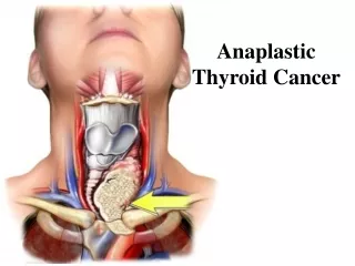
Anaplastic cancer • Peak onset age 65 and older Very rare in young patients • Males more common than females by 2 to 1 ratio • Undifferentiated • May arise many years (>20) following radiation exposure. • Neck mass usually large, diffuse, and very hard • Rapidly growing, often inoperable, highly recurrent
Invade locally, metastasize both locally and distantly (to lungs or bones) • Cervical metastasis are present in the vast majority (over 90%) of cases at the time of diagnosis. • Mean survival 6 months • Often requires the patient to get a tracheostomy to maintain their airway.
STAGING OF THYROID CANCER In differentiated thyroid carcinoma, several classification and staging systems have been introduced. However, no clear consensus has emerged favoring any one method over another • AMES system/AGES System/GAMES system • TNM system • MACIS system • University of Chicago system • Ohio State University system • National Thyroid Cancer Treatment Cooperative Study (NTCTCS)
TNM Staging • Primary tumor (T) (All categories may be subdivided into (a) solitary tumor or (b) multifocal tumor.) TX: Primary tumor cannot be assessed T0: No evidence of primary tumor T1: Tumor ≤ 2 cm, limited to the thyroid T2: Tumor > 2 cm but ≤4 cm, limited to the thyroid T3: Tumor > 4 cm limited to the thyroid or any tumor with minimal extrathyroid extension (e.g., extension to sternothyroid muscle or perithyroid soft tissues)
T4a: Tumor of any size extending beyond the thyroid capsule to invade subcutaneous soft tissues, larynx, trachea, esophagus, or recurrent laryngeal nerve • T4b: Tumor invades prevertebral fascia or encases carotid artery or mediastinal vessels All anaplastic carcinomas are considered T4 tumors. • T4a: Intrathyroidal anaplastic carcinoma—surgically resectable • T4b: Extrathyroidal anaplastic carcinoma—surgically unresectable
Regional lymph nodes (N) (Regional lymph nodes are the central compartment, lateral cervical, and upper mediastinal lNs) • NX: Regional lymph nodes cannot be assessed • N0: No regional lymph node metastasis • N1: Regional lymph node metastasis • N1a: Metastasis to level VI (pretracheal, paratracheal, and prelaryngeal/Delphian on the cricothyroid membrane (precricoid) lymph nodes) • N1b: Metastasis to unilateral or bilateral cervical or superior mediastinal lymph nodes
Distant metastases (M) • MX: Distant metastasis cannot be assessed • M0: No distant metastasis • M1: Distant metastasis
AJCC Stage Groupings Papillary or follicular thyroid cancer • Younger than 45 years • Stage I • Any T, any N, M0 • Stage II • Any T, any N, M1 • Age 45 years and older • Stage I • T1, N0, M0 • Stage II • T2, N0, M0 • Stage III • T3, N0, M0 • T1, N1a, M0 • T2, N1a, M0 • T3, N1a, M0
Papillaryorfollicularthyroidcancer Age 45 years and older • Stage I • T1, N0, M0 • Stage II • T2, N0, M0 • Stage III • T3, N0, M0 • T1, N1a, M0 • T2, N1a, M0 • T3, N1a, M0 • Stage IVA • T4a, N0, M0 • T4a, N1a, M0 • T1, N1b, M0 • T3, N1b, M0 • T2, N1b, M0 • T4a, N1b, M0 • Stage IVB • T4b, any N, M0 • Stage IVC • Any T, any N, M1
Stage IVA • T4a, N0, M0 • T4a, N1a, M0 • T1, N1b, M0 • T2, N1b, M0 • T3, N1b, M0 • T4a, N1b, M0 • Stage IVB • T4b, any N, M0 • Stage IVC • Any T, any N, M1 Medullary thyroid cancer • Stage I • T1, N0, M0 • Stage II • T2, N0, M0 • Stage III • T3, N0, M0 • T1, N1a, M0 • T2, N1a, M0 • T3, N1a, M0
Anaplastic thyroid cancer • All anaplastic carcinomas are considered stage IV. • Stage IVA • T4a, any N, M0 • Stage IVB • T4b, any N, M0 • Stage IVC • Any T, any N, M1
University of Chicago system: • Class I—disease limited to the thyroid gland • Class II—lymph node involvement • Class III—extrathyroidal invasion • Class IV—distant metastases.
PROGNOSIS Prognostic schemes: GAMES scoring (PAPILLARY & FOLLICULAR CANCER) • G Grade • A Age of patient when tumor discovered • M Metastases of the tumor (other than Neck LN) • E Extent of primary tumor • S Size of tumor (>5 cm) • The patient is then placed into a high or low risk category
Prognostic Risk Classification for Patients with Well-Differentiated Thyroid Cancer(GAMES ) Low Risk High Risk • Grade Well Differentiated Poorly Differentiated • Age <40 >40 • Mets None Regional or Distant • Extent No local extension, Capsular invasion, intrathyroidal, extrathyroidal • Sex Female Male
MACIS Scoring • Developed by the Mayo Clinic for staging. • It is known to be the most accurate predictor of a patient's outcome with papillary thyroid cancer (M = Metastasis, A = Age, I = Invasion, C = Completeness of Resection, S = Size) • MAICS Score 20 year Survival • < 6 = 99% • 6-7 = 89% • 7-8 = 56% • > 8 = 24%
Stage I and II Papillary and Follicular I-Total thyroidectomy: • Rationale? Bilateral cancers are common (30-85%) improved effectiveness for I131 ablation lowers dose needed for I131 ablation allows f/u with thyroglobulin levels decreased recurrence in all groups improved survival in high risk pts. Decreased risk of pulmonary mets • Disadvantage? higher incidence of hypoparathyroidism, but this complication may be reduced when a small amount of tissue remains on the contralateral side.
II-Lobectomy: • Rationale? Most patients are low risk and excellent prognosis Role of adjuvant treatment not defined Complications of Total Occult multicentric tumor not clinically significant Most local recurrences treated with surgery Excellent outcome with lobectomy in low risk patients • Disadvantage? • approximately 5% to 10% of patients will have a recurrence
Indications for total Thyroidectomy OR lobectomy: (all present) • Age 15 y - 45 y • No prior radiation • No distant metastases • No cervical lymph node metastases • No extrathyroidal extension • Tumor < 4 cm in diameter • No aggressive variant
When complete total thyroidectomy after lobectomy: • Aggressive variant • Macroscopic multifocal disease • Positive isthmus margins • Cervical lymph node metastases • Extrathyroidal extension Aggressive=Tall cell, columnar cell, insular, oxyphilic, or poorly differentiated features
Node removal ? • Selective node removal can be performed, and radical neck dissection is usually not required. • This results in a decreased recurrence rate, but has not been shown to improve survival.
Thyroid carcinoma after lobectomy for benign lesions • III- follow up: • Negative margins • No contralateral lesion • < 1 cm in diameter • No suspicious lymph node I-Completion of thyroidectomy: • > 4 cm • Positive margins • Extra-thyroidal invasion (T3 or T4( II- Completion of Thyroidectomy or follow up: • Clinically suspicious lymph node, contralateral lesion, or perithyroidal node • Aggressive variant • Macroscopic multifocal disease • ≥1 cm in diameter
POSTSURGICAL EVALUATION AFTER THYROIDECTOMY I-No gross Residual Disease in neck: • Follow up (TSH + thyroglobulin measurement + antithyroglobulin antibodies) II- Gross Residual Disease in neck: • Resectable >>>>>>>> Surgery • Irresectable >>>>>>>> Total body radioiodine scan: • Inadequate uptake >>>>>>RT • Adequate uptake >>>>> Radioiodine treatment or RT • No scan performed >>>>>Radioiodine treatment or RT • Total body radioiodine scan is done after adequate TSH stimulation (thyroid withdrawal or recombinant rhTSH stimulation)
Postoberative I131? a postoperative course of therapeutic (ablative) doses of I131 results in a decreased recurrence rate among high-risk patients with papillary and follicular carcinomas. Indications: (any present) • Age < 15 y or > 45 y • Radiation history • Known distant metastases • Bilateral nodularity • Extrathyroidal extension • Tumor > 4 cm in diameter • Cervical lymph node metastases • Aggressive variant
Pretherapy whole body iodine scan: • If performed, a pretherapy scan should use a low dose of 131I (1 to 5 mCi) or 123I. • To detect residual thyroid tissue, thyroid cancer, and metastatic foci • To reduce the potential for sublethal radiation stunning of thyroid tissue that prevents optimal uptake of future 131I therapy. • Stunning is defined as a reduction in uptake of the 131I therapy dose induced by a pretreatment diagnostic dose
Dose of RAI • The dosing of 131I for ablation is somewhat controversial. • Low-dose ablation with less than 30mCi administered on an outpatient basis: • For low-risk young patients • High-dose ablation with100 to 200 mCi • For high-risk patients • 300mCi • For all patients with metastatic disease that treated with repeated therapeutic doses of 131I
Replacement therapy? • Postoperative treatment with exogenous thyroid hormone in doses sufficient to suppress thyroid-stimulating hormone (TSH) with development of thyrotoxic manifestations; decreases incidence of recurrence. • Administration of Thyroid Hormone • To suppress TSH and growth of any residual thyroid • To maintain patient euthyroid • Maintain TSH level 0.1uU/ml in low risk pts • Maintain TSH Level < 0.1uU/ml in high risk pts
Stage III Papillary and Follicular A. Surgery • Total thyroidectomy plus removal of involved lymph nodes or other sites of extrathyroid disease. B. Adjuvant therapy • I131 ablation following total thyroidectomy if the tumor demonstrates uptake of this isotope. • External-beam radiation therapy if I131 uptake is minimal • Replacement therapy for all patients.
Stage IV Papillary and Follicular • Adequate uptake of I131 • I131 • Inadequate uptake or not sensitive to I131 • Localized lesions • Radiation therapy • Resection of limited metastases don't uptake of I131. • Disseminated disease • TSH suppression with thyroxine is effective. • Chemotherapy has been reported to produce occasional complete responses of long duration. • Clinical trials testing new approaches to this disease.
Medullary Thyroid Cancer treatment • Thyroidectomy: • total thyroidectomy + routine central and bilateral modified neck dissections ..Why? • External radiation therapy: • palliation of locally recurrent tumors, without evidence that it provides any survival advantage. • Radioactive iodine has no place in the treatment of patients with MTC. • Palliative chemotherapy: • Palliative chemotherapy has been reported to produce occasional responses in patients with metastatic disease. • No single drug regimen can be considered standard. • Some patients with distant metastases will experience prolonged survival and can be observed until they become symptomatic.