Download
1 / 72
720 likes | 892 Views
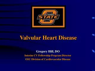
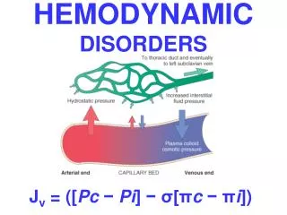
Valvular Disorders & Infecive Endocarditis. Normal Valve Function. Prevent backward flow of blood Permit forward flow of blood. Abnormal valve function. Allows backward flow valve is “leaky;” “regurgitant;” “incompetent” Reduces cardiac output while increasing workload

E N D
Normal Valve Function • Prevent backward flow of blood • Permit forward flow of blood
Abnormal valve function • Allows backward flow • valve is “leaky;” “regurgitant;” “incompetent” • Reduces cardiac output while increasing workload • results in inefficient pumping; greater volume of blood needs to be pumped with each beat to maintain cardiac output • “volume load” • Typically causes dilatation of the cardiac chamber • Backwards jet causes turbulence that is audible as murmur
Abnormal valve function • Prevents forward flow • valve does not open well • Greek stenōsis, a narrowing • Reduces cardiac output while increasing workload • Heart must develop more pressure to move blood • “pressure load” • usually results in hypertrophy of proximal (“upstream”) chamber (LA in MS, LV in AS) • acceleration of blood through tight valve causes turbulence that is audible as a murmur
Valvular Aortic Stenosis • failure of valve to open normally during systole, requiring LV to develop excess pressure to overcome increased resistance • pressure gradient between LV and aorta may be as much as 100 mm Hg • causes concentric hypertrophy • symptoms of exertional chest pain, syncope, dyspnea • mandate valve replacement to prevent sudden death
Grades of AS • Normal valve area 3-4 cm2 • Mild AS >1.5 cm2 • Moderate >1.0 cm2 • Severe AS when area ¼ normal • <1 cm2 for large person • <0.75 cm2 for normal person
Clinical Presentation of Aortic Stenosis • Cardinal symptoms: • Angina • Occurs in >50% of patients, not sensitive due to prevalence of CAD • Reduced coronary flow reserve • Increased demand-high afterload • Syncope(exertional pre-syncope) • Fixed cardiac output • Vasodepressor response • CHF • Sudden cardiac death rare, <1% per year • Other signs of LV failure • Diastolic & systolic dysfunction • In earlier stages, AS presentation more subtle • Dyspnea • Decreased exercise tolerance • Rarely, AS diagnosed in the setting of GI bleeding • Heyde’s syndrome • Bleeding caused by AVM • Concurrent AS occurs at prevalence rate of 15-25% • Associated with an acquired von Willebrand syndrome due to disruption of vW multimers through a diseased AV
Physical Findings S1 S2 S1 S2 Mild-Moderate Severe
Symptomatic AS- management • NO SAFE MEDICAL RX for Severe AS • Physical diagnosis straight forward • systolic crescendo-decrescendo murmur • loudest in aortic area usually (sometimes apex) • radiates to carotids • LV hypertrophy associated with gallop (S4) • Signs of critical AS • carotid upstrokes small and delayed in severe AS • loss of aortic component of S2 • late peaking murmur
Management of Aortic Stenosis • Prognosis in asymptomatic disease excellent • Conservative approach with monitoring for symptoms recommended • When severe stenosis present- • 38% of asymptomatic patients develop symptoms within 2 years • 79% are symptomatic within 3 years • Once symptoms occur, AVR needed • LV dysfunction and severe AS have increased perioperative mortality with AVR • But outcomes still favorable with surgery • Nitroprusside may transiently improve cardiac function as a bridge to valve replacement • Does not supplant AVR in symptomatic patients
Aortic Valve Replacement • Prophylatic AVR in asymptomatic patients not routinely performed due to surgical risks • Thromboembolism, bleeding associated with anticoagulation, prosthetic valve dysfunction, and endocarditis • Occurs at a rate of 2-3% annually • Only should be considered: • If other cardiac surgery (such as CABG) planned • Severe LVH or systolic dysfunction • Women contemplating pregnancy • Patients remote from health care • Surgical valve replacement with operative morbidity and mortality of 10% • Percutaneous balloon aortic valvotomy rarely used
Any conditions resulting in incompetent aortic leaflets Congenital Bicuspid valve Aortopathy Cystic medial necrosis Collagen disorders (e.g. Marfan’s) Ehler-Danlos Osteogenesis imperfecta Pseudoxanthoma elasticum Acquired Rheumatic heart disease Dilated aorta (e.g. hypertension..) Degenerative Connective tissue disorders E.g. ankylosing spondylitis, rheumatoid arthritis, Reiter’s syndrome, Giant-cell arteritis ) Syphilis (chronic aortitis) Acute AI: aortic dissection, infective endocarditis, trauma Aortic Regurgitation: Etiology
Aortic Regurgitation: Symptoms • Dyspnea, orthopnea, PND • Chest pain. • Nocturnal angina >> exertional angina • ( diastolic aortic pressure and increased LVEDP thus coronary artery diastolic flow) • With extreme reductions in diastolic pressures (e.g. < 40) may see angina
Quincke’s sign: capillary pulsation Corrigan’s sign: water hammer pulse Bisferiens pulse (AS/AR > AR) De Musset’s sign: systolic head bobbing Mueller’s sign: systolic pulsation of uvula Durosier’s sign: femoral retrograde bruits Traube’s sign: pistol shot femorals Hill’s sign:BP Lower extremity >BP Upper extremity by > 20 mm Hg - mild AR > 40 mm Hg – mod AR > 60 mm Hg – severe AR Peripheral Signs of Severe Aortic Regurgitation
Aortic Regurgitation: Physical Exam • Widened pulse pressure • Systolic – diastolic = pulse pressure • High pitched, blowing, decrescendo diastolic murmur at LSB • Best heard at end-expiration & leaning forward • Hands & Knee position S1S2 S1
Aortic Regurgitation: Natural History Asymptomatic %/Y • Normal LV function (~good prognosis) • Progression to symptoms or LV dysfunction < 6 • Progression to asymptomatic LV dysfunction < 3.5 • 75% 5-year survival • Sudden death < 0.2 • Abnormal LV function • Progression to cardiac symptoms 25 • Symptomatic (Poor prognosis) • Mortality > 10 TX: Medical Surgery BEFORE LV dysfunction
Aortic Regurgitation • Loss of cardiac output backwards from aorta into LV • congenital, endocarditis, age, aortic disease, collagen vascular, syphillis • Early diastolic, decrescendo murmur best heard at LLSB with diaphragm • subtle, have pt lean forward, breathe out • associated with wide pulse pressure
Mitral Stenosis • Almost always rheumatic in origin • >40% of cases of RHD result in mitral stenosis • Women affected more than men (2:1) • Presentation 20-40 years after the initial episode of rheumatic fever • If infected at a young age, latent period is a few years • Murmur may be subtle, but high flow states cause increased pressure gradient, pulmonary edema • classic presentation is during vaginal delivery. Tachycardia, straining, volume increase cause pulmonary edema • Patients eventually have exertional dyspnea, atrial fibrillation (often with thromboembolism), chest pain
Mitral Stenosis • Turbulent, high velocity flow occurs during diastole • murmur is therefore a DIASTOLIC, low frequency rumble heard at apex with stethoscope bell, patient in L lateral decubitus • requires quiet concentration, palpate carotid to time systole/diastole • Always look for MS in patient with new Atrial fibrillation • rate control, anticoagulation crucial
Mitral Stenosis Pathophysiology • Normal valve area: 4-6 cm2 • Mild mitral stenosis: • MVA 1.5-2.5 cm2 • Minimal symptoms • Mod mitral stenosis • MVA 1.0-1.5 cm2 usually does not produce symptoms at rest • Severe mitral stenosis • MVA < 1.0 cm2
Fatigue Palpitations Cough SOB Left sided failure Orthopnea PND Palpitation AFib Systemic embolism Pulmonary infection Hemoptysis Right sided failure Hepatic Congestion Edema Worsened by conditions that cardiac output. Exertion,fever, anemia, tachycardia, Afib, intercourse, pregnancy, thyrotoxicosis Mitral Stenosis Symptoms
Mitral Stenosis Physical Exam • First heart sound (S1) is accentuated and snapping • Opening snap (OS) after aortic valve closure • Low pitch diastolic rumble at the apex • Pre-systolic accentuation (esp. if in sinus rhythm) S1 S2 OS S1
Management of Mitral Stenosis • Atrial fibrillation • Prevalence >30% in symptomatic patients and associated with poorer long term outcome • Warfarin indicated: • In patients with AF and MS • Patients without history of AF but with MS and embolic CVA • In patients with prior history of AF who have mitral valve surgery, decreased postoperative AF observed if MAZE performed concominantly
MS Mortality • Minimal sxs >80% 10 year survival • Limiting sxs, <15% 10 year survival • Untreated patients • 60-70% progressive pulmonary edema • 20-30% systemic embolism • 10% pulmonary embolism • 1-5% endocarditis/infection
Mitral Regurgitation • Incompetent mitral valve allows loss of stroke volume back into LA • Mitral valve prolapse most common cause • rheumatic disease and endocarditis • PE much less subtle than MS • loud pan-SYSTOLIC murmur, loudest at apex and radiating into axilla • typically soft S1 • S2 obscured by murmur • presence of S3 suggests severe MR
Valvular-leaflets Myxomatous MV Disease Rheumatic Endocarditis Congenital-clefts Chordae Fused/inflammatory Torn/trauma Degenerative IE Annulus Calcification, IE (abcess) Papillary Muscles CAD (Ischemia, Infarction, Rupture) HCM Infiltrative disorders LV dilatation & functional regurgitation Trauma Mitral Regurgitation: Etiology
MRSymptoms • Similar to MS • Dyspnea, Orthopnea, PND • Fatigue • Pulmonary HTN, right sided failure • Hemoptysis • Systemic embolization in A Fib
Mitral Insufficiency: Physical Exam • Fixed mitral regurgitation • Mitral valve prolapse S1 S2 S1 S1 C S2
ECG: LA enlargement Afib LVH (50% pts. With severe MR) RVH (15%) Combined hypertrophy (5%) CXR: LV LA pulmonary vascularity CHF Ca++ MV/MAC Recognizing Mitral Regurgitation
MR Treatment • No medical therapy • Most difficult clinically • By the time symptoms occur, it may be too late • Drop in EF or development of atrial fibrillation enough to justify surgery
Mitral Valve Prolapse : Epidemiology • Affects 5-10% of population • Most common cause of isolated severe MR • Females >> males; Ages of 14 and 30years • Strong hereditary component (? Autosomal Dominant) • 2º to failure of apposition/coaptation of the anterior and posterior mitral valve leaflets. • Results form diverse pathologic conditions, but cause is unknown in a majority of pts
Mitral Valve Prolapse: Symptoms • Majority are asymptomatic for entire life • Palpitations • Chest pain (atypical). • Often substernal, prolonged, poorly related to exertion, and rarely resembles typical angina • Syncope
Mitral Valve Prolapse: Physical Exam • Most important finding: mid late systolic click. • Acute tensing of the mitral valve chordae • Variable murmurs: • high pitched late systoliccrescendo-decrescendo murmur, • Occasionally “whooping” or “honking” at the apex S1 C S2
Mitral Valve Prolapse: Complications • Arrhythmias (Usually PVC, PSVT>>VT) • Transient cerebral ischemic (embolic – rare) • Infective endocarditis (if assoc w/ MR) • Sudden death (rare)
Tricuspid and Pulmonic Valve Disorders Etiology/Pathophysiology/Manifestations Tricuspid stenosis (more common than regurgitation) Result in R. atrial enlargement > inc. systemic venous pressure > atrial fibrillation, peripheral edema, ascites, etc. Found mostly in rheumatic heart disease, IV drug users Pulmonic stenosis Result in R. ventricular hypertension and hypertrophy Fatigue , loud midsystolic murmur Uncommon valve disorders
Tricuspid Valve Disease • Tricuspid stenosis is rare • Associated with rheumatic heart disease • TR usually occurs secondary to: • Pulmonary hypertension • RV chamber enlargement with annular dilatation • Endocarditis (associated with IV drug use) • Injury following pacer lead placement • Other secondary causes: carcinoid, radiation therapy, anorectic drug use, and trauma • Primary causes: Marfan’s syndrome and congenital disorders such as Ebstein’s anomaly and AV canal malformation • Echo is diagnostic in most cases
Tricuspid Regurgitation • Severe tricuspid regurgitation is difficult to treat and carries a poor overall clinical outcome • Symptoms are manifestations of systemic venous congestion • Ascites • Pedal edema • Surgical intervention usually considered if other cardiac surgery planned • Surgical options include valvular annuloplasty or replacement • If replacement planned, bioprosthetic valve preferred
Other Valve disorders: NBTE: Non bacterial thrombotic… Thrombus on valves – Hypercoag., DIC, Malignancy, etc. May cause strokes, sec. bacterial infection. Libman-Sacks: Sterile Immune complex vegetations SLE. Carcinoid Heart Disease: Carcinoid tum, 5HT, seratonins etc.. Endocardial fibrosis
Valvular Disease • Rheumatic fever • Regurgitation frequently present acutely • Long term predominant effect is stenosis • Endocarditis causes regurgitation • Patients with valve dz should take antibiotics prior to dental work to prevent endocarditis • All patients with symptomatic valvular disease (i.e. dyspnea, chest pain, syncope) need to be evaluated for surgical correction • Some asymptomatic subjects also need correction “before it’s too late”
Valvular DiseaseGeneral Principles • Left sided valvular disease more prone to cause serious hemodynamic problems • Regurgitation causes volume overload- eccentric hypertrophy (dilatation) • Stenotic lesions cause pressure overload on proximal chamber- concentric hypertrophy (thickened walls) • Stenotic lesions cause symptoms sooner than regurgitant lesions but respond to therapy better
Common Murmurs and Timing Systolic Murmurs • Aortic stenosis • Mitral insufficiency • Mitral valve prolapse • Tricuspid insufficiency Diastolic Murmurs • Aortic insufficiency • Mitral stenosis S1 S2 S1
Surgical Intervention *Not all types valve disease require surgical intervention Valvuloplasty-general term valve repair, invasive/non-invasive methods Percutaneous balloon valvuloplasty (non-invasive) Surgery Open commissurotomy- open stenotic valves Annuloplasty- repair of valve’s outer ring-used for stenosis, regurgitant valve Valve Replacement Mechanical-need anticoagulant Biologic-only last about 15 years
Prosthetic Valve Complications • Common complications include: • Structural valve deterioration • Valve thrombosis • Embolism • Bleeding • Endocarditis • Endocarditis prophylaxis required for patients with all types of prosthetic valves • Suspect valve dehiscence or dysfunction in: • Acute CHF in the immediate postop period • New cardiac symptoms • Embolic phenomena • Hemolytic anemia • New murmurs • TEE is the diagnostic procedure of choice • Postop TTE should be done 2-3 months after surgery
1997 American Heart Assoc. Guidelines: Endocarditis Prophylaxis Recommended:1997 High-risk category Prosthetic cardiac valves, including bioprosthetic and homograft valves Previous BE Complex cyanotic congenital heart disease (eg, single ventricle states, transposition of the great arteries, Tetralogy of Fallot) Surgically constructed systemic pulmonary shunts or conduits Moderate-risk category :1997 Most other congenital cardiac malformations (other than above and below) Acquired valvar dysfunction (eg,RHD) HOCM MVP with valvar regurgitation and/or thickened leaflets
Endocarditis Prophylaxis Not Recommended: Negligible-risk category : 1997 (No greater risk than the general population) Isolated secundum atrial septal defect Surgical repair ofASD,VSD, or (without residua beyond 6 mo) Previous CABG MVP without valvar regurgitation1 Physiologic, functional, or innocent heart murmurs1 Previous Kawasaki disease without valvar dysfunction Previous rheumatic fever without valvar dysfunction Cardiac pacemakers (intravascular and epicardial) and implanted defibrillators
2007: Who gets prophylaxis? Only patients with the highest risk of adverse outcomes (heart failure, surgery, death) from endocarditis: 1. Prosthetic cardiac valve 2. Previous IE 3. Cardiac transplant recipients who develop cardiac valvulopathy 4. Congenital Heart Disease