Download
1 / 30
320 likes | 586 Views
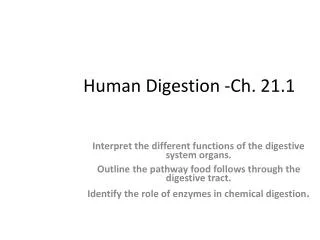
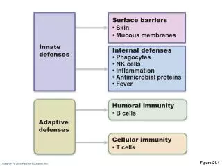
Surface barriers • Skin • Mucous membranes. Innate defenses. Internal defenses • Phagocytes • NK cells • Inflammation • Antimicrobial proteins • Fever. Humoral immunity • B cells. Adaptive defenses. Cellular immunity • T cells. Figure 21.1. Inflammatory Response.

E N D
Surface barriers • Skin • Mucous membranes Innate defenses Internal defenses • Phagocytes • NK cells • Inflammation • Antimicrobial proteins • Fever Humoral immunity • B cells Adaptive defenses Cellular immunity • T cells Figure 21.1
Inflammatory Response • Macrophages and epithelial cells of boundary tissues bear Toll-like receptors (TLRs) • TLRs recognize specific classes of infecting microbes • Activated TLRs trigger the release of cytokines that promote inflammation
Phagocyte Mobilization • Steps for phagocyte mobilization • Leukocytosis: release of neutrophils from bone marrow in response to leukocytosis-inducing factors from injured cells • Margination: neutrophils cling to the walls of capillaries in the inflamed area • Diapedesis of neutrophils • Chemotaxis: inflammatory chemicals (chemotactic agent) promote positive chemotaxis of neutrophils
Innate defenses Internal defenses Tissue injury Release of chemical mediators (histamine, complement, kinins, prostaglandins, etc.) Release of leukocytosis- inducing factor Leukocytosis (increased numbers of white blood cells in bloodstream) Vasodilation of arterioles Increased capillary permeability Attract neutrophils, monocytes, and lymphocytes to area (chemotaxis) Leukocytes migrate to injured area Local hyperemia (increased blood flow to area) Capillaries leak fluid (exudate formation) Margination (leukocytes cling to capillary walls) Initial stimulus Physiological response Signs of inflammation Diapedesis (leukocytes pass through capillary walls) Leaked clotting proteins form interstitial clots that wall off area to prevent injury to surrounding tissue Leaked protein-rich fluid in tissue spaces Result Phagocytosis of pathogens and dead tissue cells (by neutrophils, short-term; by macrophages, long-term) Heat Redness Pain Swelling Temporary fibrin patch forms scaffolding for repair Locally increased temperature increases metabolic rate of cells Possible temporary limitation of joint movement Pus may form Area cleared of debris Healing Figure 21.3
Phagocyte Mobilization • Steps for phagocyte mobilization • Leukocytosis: release of neutrophils from bone marrow in response to leukocytosis-inducing factors from injured cells • Margination: neutrophils cling to the walls of capillaries in the inflamed area • Diapedesis of neutrophils • Chemotaxis: inflammatory chemicals (chemotactic agent) promote positive chemotaxis of neutrophils
Innatedefenses Internaldefenses Inflammatorychemicalsdiffusingfrom theinflamed siteact as chemotacticagents. Capillary wall Basementmembrane Endothelium 1 Leukocytosis.Neutrophils enter bloodfrom bone marrow. Figure 21.4, step 1
Innatedefenses Internaldefenses Inflammatorychemicalsdiffusingfrom theinflamed siteact as chemotacticagents. Capillary wall Basementmembrane Endothelium 1 2 Leukocytosis.Neutrophils enter bloodfrom bone marrow. Margination.Neutrophils clingto capillary wall. Figure 21.4, step 2
Innatedefenses Internaldefenses Inflammatorychemicalsdiffusingfrom theinflamed siteact as chemotacticagents. Capillary wall Basementmembrane Endothelium 1 2 3 Leukocytosis.Neutrophils enter bloodfrom bone marrow. Margination.Neutrophils clingto capillary wall. Diapedesis.Neutrophils flatten andsqueeze out of capillaries. Figure 21.4, step 3
Innatedefenses Internaldefenses Inflammatorychemicalsdiffusingfrom theinflamed siteact as chemotacticagents. 4 Chemotaxis.Neutrophilsfollow chemicaltrail. Capillary wall Basementmembrane Endothelium 1 2 3 Leukocytosis.Neutrophils enter bloodfrom bone marrow. Margination.Neutrophils clingto capillary wall. Diapedesis.Neutrophils flatten andsqueeze out of capillaries. Figure 21.4, step 4
Adaptive Defenses • Adaptive immune response • Is specific • Is systemic • Has memory • Two separate overlapping arms • Humoral (antibody-mediated) immunity • Cellular (cell-mediated) immunity
Antigens • Substances that can mobilize the adaptive defenses and provoke an immune response • Most are large, complex molecules not normally found in the body (nonself)
Antigenic Determinants • Parts of antigen that are immunogenic • Antibodies and lymphocyte receptors bind to them
Antigen- binding sites Antigenic determinants Antibody A Antigen Antibody B Antibody C Figure 21.7
Haptens (Incomplete Antigens) • Small molecules (peptides, nucleotides, and hormones) • Not immunogenic by themselves • Are immunogenic when attached to body proteins • Cause the immune system to mount a harmful attack • Examples: poison ivy, animal dander, detergents, and cosmetics
Self-Antigens: MHC Proteins • Proteins (self-antigens) on cell surface of s • Example: MHC proteins • Coded for by genes of major histocompatibility complex (MHC) and unique
MHC Proteins • Classes • Class I MHC proteins, on most all cells • Class II MHC proteins, on certain cells in immune response
Cells of the Adaptive Immune System • Two types of lymphocytes • B lymphocytes (B cells)—humoral immunity • T lymphocytes (T cells)—cell-mediated immunity • Antigen-presenting cells (APCs) • Do not respond to specific antigens • Play essential auxiliary roles in immunity
Red bone marrow: site of lymphocyte origin Humoral immunity Adaptive defenses Cellular immunity Primary lymphoid organs: site of development of immunocompetence as B or T cells Immature lymphocytes Red bone marrow Secondary lymphoid organs: site of antigen encounter, and activation to become effector and memory B or T cells 1 Lymphocytes destined to become T cells migrate (in blood) to the thymus and develop immunocompetence there. B cells develop immunocompetence in red bone marrow. Thymus Bone marrow 2 Immunocompetent but still naive lymphocytes leave the thymus and bone marrow. They “seed” the lymph nodes, spleen, and other lymphoid tissues where they encounter their antigen. Lymph nodes, spleen, and other lymphoid tissues 3 Antigen-activated immunocompetent lymphocytes (effector cells and memory cells) circulate continuously in the bloodstream and lymph and throughout the lymphoid organs of the body. Figure 21.8
T Cells • T cells mature in the thymus under negative and positive selection pressures • Positive selection • Selects T cells capable of binding to self-MHC proteins (MHC restriction) • Negative selection • Prompts apoptosis of T cells that bind to self-antigens displayed by self-MHC • Ensures self-tolerance
Adaptive defenses Cellular immunity Positive selection: T cells must recognize self major histocompatibility proteins (self-MHC). Antigen- presenting thymic cell Developing T cell Failure to recognize self-MHC results in apoptosis (death by cell suicide). MHC T cell receptor Self-antigen Recognizing self-MHC results in MHC restriction—survivors are restricted to recognizing antigen on self-MHC. Survivors proceed to negative selection. Negative selection: T cells must notrecognize self-antigens. Recognizing self-antigen results in apoptosis. This eliminates self-reactive T cells that could cause autoimmune diseases. Failure to recognize (bind tightly to) self-antigen results in survival and continued maturation. Figure 21.9
Antigen Receptor Diversity • Lymphocytes make up to a billion different types of antigen receptors • Coded for by ~25,000 genes • Gene segments are shuffled by somatic recombination • Genes determine which foreign substances the immune system will recognize and resist
Antigen-Presenting Cells (APCs) • Engulf antigens • Present fragments of antigens to T cells • Major types • Dendriticin connective tissues and epidermis • Macrophages in connective tissues and lymphoid organs • B cells
Macrophages and Dendritic Cells • Present antigens and activate T cells • Macrophages mostly remain fixed in the lymphoid organs • Dendritic cells internalize pathogens and enter lymphatics to present the antigens to T cells in lymphoid organs • Activated T cells release chemicals that • Prod macrophages to become insatiable phagocytes and to secrete bactericidal chemicals
Adaptive defenses Humoral immunity Antigen Primary response (initial encounter with antigen) Antigen binding to a receptor on a specific B lymphocyte (B lymphocytes with non-complementary receptors remain inactive) Proliferation to form a clone Activated B cells Plasma cells (effector B cells) Memory B cell— primed to respond to same antigen Secreted antibody molecules Figure 21.11 (1 of 2)
Clonal Selection • B cell is activated when antigens bind to its surface receptors and cross-link them • Receptor-mediated endocytosis of cross-linked antigen-receptor complexes occurs • Stimulated B cell grows to form a clone of identical cells bearing the same antigen-specific receptors(T cells are usually required to help B cells achieve full activation)
Fate of the Clones • Secreted antibodies • Circulate in blood or lymph • Bind to free antigens • Mark the antigens for destruction
Immunological Memory • Primary immune response • Occurs on the first exposure to a specific antigen • Lag period: three to six days • Peak levels of plasma antibody are reached in 10 days • Antibody levels then decline
Adaptive defenses Humoral immunity Antigen Primary response (initial encounter with antigen) Antigen binding to a receptor on a specific B lymphocyte (B lymphocytes with non-complementary receptors remain inactive) Proliferation to form a clone Activated B cells Plasma cells (effector B cells) Memory B cell— primed to respond to same antigen Secreted antibody molecules Subsequent challenge by same antigen results in more rapid response Secondary response (can be years later) Clone of cells identical to ancestral cells Plasma cells Secreted antibody molecules Memory B cells Figure 21.11
Secondary immune response to antigen A is faster and larger; primary immune response to antigen B is similar to that for antigen A. Primary immune response to antigen A occurs after a delay. Anti- bodies to B Anti- bodies to A Second exposure to antigen A; first exposure to antigen B First exposure to antigen A Time (days) Figure 21.12
Humoral immunity Active Passive Naturally acquired Naturally acquired Artificially acquired Artificially acquired Infection; contact with pathogen Antibodies pass from mother to fetus via placenta; or to infant in her milk Vaccine; dead or attenuated pathogens Injection of immune serum (gamma globulin) Figure 21.13